Welcome to the Dr Hugo Kitchen Knowledge Library
Moles & Skin Lesions
Few things cause more anxiety than discovering a new mole, noticing a changing skin lesion, or feeling a lump beneath the skin.
For many people, the first thought is, “Could this be skin cancer?” That worry is entirely understandable, and it is one of the commonest reasons patients seek medical advice.
After nearly four decades of assessing and treating skin lesions, I have learnt that knowledge is one of the most powerful medicines we can offer. Most skin lesions prove to be completely harmless, yet uncertainty can create considerable anxiety. My aim is not simply to diagnose and treat skin conditions, but to help you understand what you are seeing and why it may or may not be a cause for concern.
This Knowledge Hub has been written to answer the questions I hear most frequently in clinic. It explains how to recognise common skin lesions, when you should seek medical advice, what happens during an assessment, and the treatment options available for a wide variety of benign and potentially serious conditions.
Whether you are concerned about a mole, a skin tag, a cyst, a lipoma, a wart, xanthelasma or another skin lesion, I hope this guide provides reassurance, practical advice and a clearer understanding of your skin.
Of course, no website can replace a face-to-face medical assessment. Every person is different, and every skin lesion deserves to be considered in the context of the individual. If you remain concerned about any lesion, or simply want the reassurance of an expert opinion, I would always encourage you to seek professional assessment.
My philosophy has always been simple:
Educate first. Reassure where appropriate. Investigate when necessary. Treat with care.
I hope you find this Knowledge Hub both informative and reassuring.
Dr Hugo Kitchen
⸻
Contents
- Is My Mole Cancer?
- What Exactly Is a Mole?
- The ABCDE Guide to Melanoma
- Mole Mapping Explained
- What Happens During a Mole Assessment?
- Should I Remove My Mole?
- Skin Cancer Prevention
- Seborrhoeic Keratoses
- Skin Tags
- Skin Cysts
- Lipomas
- Warts & Verrucae
- Xanthelasma
Is My Mole Cancer ?
Removal of skin tags, warts and moles in Warwickshire
The removal of moles is a simple and safe procedure when carried out by a trained and experienced clinician. The procedure can be carried out to remove moles or skin tags that are unsightly or causing irritation. Procedures are carried out under local aesthetic and are virtually painless.
Removals carried out at our Warwickshire clinic delivers excellent results with little or no scarring, even for facial treatments. Dr Hugo Kitchen is highly skilled and extremely experienced having carried out hundreds of skin tag and mole removal procedures.
We also provide a full follow up consultation and send suspicious moles to a histologist for fast diagnosis. The whole procedure can be completed in one visit, and in many cases takes no more than one hour. We also offer Mole-mapping and Artificial Intelligence driven Dermoscopy and can store your mole photographic data for the future comparison if there has been a change.
Understanding Moles – The Science Without the Scare
Before we discuss the warning signs of melanoma, it’s worth understanding what a mole actually is. I often find that once patients understand the biology, much of their anxiety disappears. If you have concerns read my guide to the ABCDE of mole watching.
A mole, or melanocytic naevus to give it its medical name, is simply a cluster of pigment-producing cells called melanocytes. These cells are responsible for making melanin, the pigment that gives our skin, hair and eyes their colour and provides some protection from ultraviolet radiation.
For reasons that are not yet completely understood, these cells sometimes gather together into small nests rather than remaining evenly distributed throughout the skin. The result is what we recognise as a mole.
Some babies are born with moles, although most appear during childhood and adolescence. It is perfectly normal for new moles to develop into your twenties and, occasionally, your thirties. Hormonal changes during puberty or pregnancy may also cause existing moles to darken slightly or become more noticeable. These changes are often entirely physiological.
Indeed, if you looked closely at the skin of ten healthy adults, you would probably find that every one of them has a unique pattern of moles. Some people have only a handful, while others may have well over one hundred. Genetics plays a major role, but lifetime sun exposure also contributes.
This is one of the reasons why there is no such thing as a “normal” mole. Moles come in an astonishing variety of colours, shapes and sizes. Some are completely flat, others are raised. Some are flesh coloured, others are light brown, dark brown or almost black. Many have tiny hairs growing through them. Contrary to popular belief, hairs growing from a mole are usually a reassuring rather than an alarming feature.
Patients often ask me, “Doctor, why has this mole suddenly appeared?”
In many cases, there is no sinister explanation whatsoever. The skin is a remarkably dynamic organ. New moles can develop naturally, particularly in younger adults. Seborrhoeic keratoses —one of the commonest benign skin growths—are frequently mistaken for new moles. Sun damage accumulated over decades can also produce lesions that patients suddenly notice one morning despite them having evolved slowly over several years.
The important point is this: finding a new mole does not automatically mean skin cancer.
Equally, having a mole for twenty or even thirty years does not guarantee that it should never be examined if it begins to change. Medicine is rarely black and white. It is about recognising patterns, identifying exceptions and knowing when further assessment is sensible.
One of the greatest misconceptions I encounter is the belief that melanoma always develops from an existing mole. In reality, many melanomas arise as entirely new pigmented lesions rather than evolving from a mole that has been present for years. That is why becoming familiar with your skin as a whole is often more valuable than obsessively monitoring one individual mole.
Another myth is that every dark mole is dangerous. Again, this is simply untrue. Some of the darkest moles I see prove to be completely benign, while occasionally a melanoma may be surprisingly pale. Colour alone tells only part of the story.
So what am I looking for when I assess a patient?
Firstly, I take a careful history. Has the lesion changed? Over what period? Is it itching, bleeding, crusting or failing to heal? Is there a personal or family history of melanoma? Has there been significant childhood sunburn? Has the patient noticed similar lesions elsewhere?
Only then do I begin the physical examination.
To the naked eye I assess symmetry, borders, colour and overall appearance. I then examine the lesion using dermoscopy—a specialised magnifying instrument with polarised light that allows me to visualise pigment networks, blood vessels and structures hidden beneath the skin surface. It is one of the most valuable advances in skin cancer diagnosis over the past few decades.
More recently, artificial intelligence has become another useful tool in clinical practice. Used responsibly, AI can compare dermoscopic images with thousands of documented lesions, offering an additional opinion that complements clinical judgement. However, I regard AI as exactly that—an assistant. It can never replace a thorough history, careful examination and the experience gained from seeing many thousands of lesions over nearly four decades.
Patients are often surprised when I tell them that I spend as much time reassuring people as I do referring them. Thankfully, the overwhelming majority of lesions I assess are benign. Sometimes all that is required is explanation, education and the confidence that comes from knowing an experienced clinician has examined the lesion carefully.
That reassurance has genuine value. Anxiety itself can become a burden, and one of my aims has always been to replace uncertainty with understanding.
Of course, there are occasions when I recommend removal or referral. If a lesion demonstrates suspicious dermoscopic features, is changing significantly or simply cannot be confidently classified as benign, then further investigation is the correct course of action. Modern medicine is at its best when it combines vigilance with common sense—not unnecessary alarm, but not false reassurance either.
Over the years I have often reflected that patients rarely remember the technical details of a consultation. What they remember is how they felt when they left the room. Did they feel listened to? Did they understand what had been explained? Were they reassured without being dismissed?
That has always been my philosophy.
Knowledge should replace fear, but knowledge delivered with kindness is even more powerful.
This article has been written to help you understand your skin-not to diagnose it. If you have any concern about a mole or skin lesion, nothing can replace a careful examination by an experienced clinician.
The Dr Hugo Kitchen Knowledge Library
1. Is My Mole Cancer?
If you’re unsure whether a lesion should actually be removed, read my guide on Should I Remove my Mole?
1. What Exactly Is a Mole?
If you’ve discovered a new mole or have become concerned about one you’ve had for many years, it helps to start with the basics. Understanding what a mole actually is often removes much of the fear that naturally accompanies the word “cancer.”
A mole, known medically as a melanocytic naevus, is one of the commonest features of normal human skin. Almost every adult has at least a few moles, while some people naturally have many dozens or even hundreds. In most cases they are entirely harmless and simply represent a normal variation in the way our skin develops.
To understand a mole, imagine looking beneath the surface of the skin.
Scattered throughout the outer layer are millions of tiny cells called melanocytes. These remarkable cells produce melanin, the natural pigment that gives our skin, hair and eyes their colour. Melanin also acts as one of the body’s natural defence mechanisms by helping to absorb some of the ultraviolet (UV) radiation from the sun.
Normally these pigment cells are spread evenly throughout the skin, rather like stars scattered across a clear night sky. Occasionally, however, a small group of melanocytes gathers together into a cluster or “nest”. When this happens, we see what we recognise as a mole.
There is nothing unusual or abnormal about this process. In fact, it is an entirely natural part of skin development.
Every Mole Is Different
One of the biggest misconceptions I encounter is the belief that there is a single “normal” appearance for a mole.
The truth is quite the opposite.
Healthy moles come in an extraordinary variety of shapes, colours and sizes.
They may be:
- Completely flat or raised above the skin.
- Smooth, slightly rough or dome-shaped.
- Light brown, dark brown, almost black or even flesh coloured.
- Perfectly round, oval or slightly irregular.
- Tiny pinhead lesions or several millimetres across.
- Hair-bearing or completely hairless.
Many patients become concerned when they notice coarse hairs growing through a mole. Surprisingly, this is usually a reassuring sign and is commonly seen in entirely benign moles.
Hairy Mole
If you’d like to understand how we use digital photography, dermoscopy and A.I. to monitor changing moles, read my guide on Mole Mapping Explained.
When Do Moles Develop?
Some moles are present from birth and are known as congenital moles, although these account for only a small proportion of all moles.
Most develop naturally during childhood, adolescence and early adulthood. Hormonal changes during puberty and pregnancy may make existing moles appear slightly darker or more prominent, and this is usually a perfectly normal response.
It is also quite common for new moles to appear during your twenties and, occasionally, into your thirties.
After the age of about forty, genuinely new moles become less common. This does not automatically mean that a new pigmented lesion is dangerous, but it does mean that it is sensible to have it examined by an experienced clinician so that the correct diagnosis can be made.
Why Do Some Moles Change?
Our skin is a living organ that is constantly adapting throughout life. Just as wrinkles gradually appear and hair changes colour, moles can also evolve over many years.
A harmless mole may slowly become raised, lose pigment, develop hairs or even gradually fade with age. These changes are often part of the normal ageing process rather than signs of disease.
Equally, not every new brown mark on the skin is actually a mole. One of the commonest reasons older patients attend my clinic is because they have developed what they believe to be a “new mole”, when in fact it proves to be a seborrhoeic keratosis—a completely benign age-related skin growth. This is one of the reasons why a proper examination is so valuable: many different skin lesions can look remarkably similar to the untrained eye.
Should I Worry About Every Mole?
The simple answer is no.
After nearly four decades examining skin lesions, I can reassure you that the overwhelming majority of moles I assess are entirely benign. Most require nothing more than explanation and reassurance.
However, medicine is never about making assumptions.
The important question is not “Do I have a mole?” but rather “Is this mole behaving differently?”
Changes in size, shape, colour, border, symptoms such as itching or bleeding, or the appearance of a lesion that looks noticeably different from your other moles should always be assessed.
Early diagnosis remains one of the most powerful tools we have against melanoma. Fortunately, most suspicious lesions prove to be something much less concerning, but knowing the difference is precisely why professional assessment is so important.
Dr Kitchen’s Clinical Pearl
One lesson I have learnt after examining many thousands of skin lesions is that patients often worry far more than they need to. In fact, one of the most rewarding parts of my work is being able to tell someone, “I’ve got some good news—that mole is entirely benign.”
Reassurance is a treatment in its own right.
Knowledge replaces fear, and understanding your own skin is the first step towards protecting it.
*The commonest diagnosis I make is not Melanoma – it’s anxiety
*Patients rarely regret having a mole checked; they often regret months of unnecessary worry before seeking advice.
*Experience teaches you that reassurance, when justified, can be as therapeutic as any procedure.
“The one question I hear almost every day.”
Few words generate as much anxiety as the phrase “skin cancer.” Every week I meet patients who have lived with a mole for years, only to become concerned after noticing a subtle change or after a friend suggests they should “get it checked.” Others arrive convinced the worst has happened because they have spent hours searching the internet, where every photograph seems to resemble melanoma.
The reality is far more reassuring.
After nearly four decades assessing skin lesions, I can tell you that the overwhelming majority of moles I examine are entirely benign. That does not mean they should be ignored, but it does mean that concern and panic are two very different things.
A mole is simply a collection of pigment-producing cells called melanocytes. Most of us have between 10 and 40 moles, and many develop during childhood and early adult life. Some slowly enlarge as we grow, while others may gradually fade with age. These are all perfectly normal changes.
The important question is not whether you have a mole, but whether that mole is behaving differently from the rest of your skin.
When I examine a patient, I don’t rely on instinct alone. I look for specific clinical features, examine the lesion using dermoscopy to reveal structures invisible to the naked eye, and increasingly use AI-assisted imaging to complement—not replace—clinical judgement. Technology is a remarkable servant but a poor master. Experience still matters.
One of the most useful concepts is what dermatologists call the “Ugly Duckling Sign.” Most of your moles look similar. The one that stands out—the one that seems different—is often the one deserving closer attention.
My advice is simple. Become familiar with your own skin. If a mole changes in size, colour, shape, becomes itchy, bleeds without injury or simply feels different, have it assessed promptly. Most changes prove harmless, but the small number that do not are far easier to treat when detected early.
Knowledge should replace fear. That is the purpose of this guide.
2. The ABCDE Guide to Melanoma
Many patients have heard of the ABCDE rule but few understand why it matters. Rather than being a checklist designed to frighten people, it is simply a practical way of recognising when a mole deserves professional assessment. In this article, we’ll explore what asymmetry, border irregularity, colour variation, diameter and evolution really mean—and why the final letter, “E” for evolution or change, is often the most important of all.
If you’ve ever searched the internet about moles or skin cancer, you have almost certainly come across something called the ABCDE Rule.
It is one of the most widely recognised tools used to help identify moles that may require further assessment. It is simple to remember, easy to apply and has undoubtedly helped many people seek medical advice at an early stage.
However, it is important to understand one thing from the outset.
The ABCDE Rule is a guide—not a diagnosis.
Many completely harmless moles may display one or even two of these features, while some early melanomas may not fulfil every criterion. Think of the ABCDE Rule as a prompt to seek professional advice rather than a way of diagnosing yourself.
A – Asymmetry
Most benign moles are reasonably symmetrical. If you were to draw an imaginary line through the middle, each half would look broadly similar.
A mole that has become noticeably asymmetrical, where one side no longer resembles the other, deserves closer attention.
That said, slight asymmetry is surprisingly common and is not, on its own, proof of skin cancer. It is simply one piece of the overall picture.
B – Border
Benign moles usually have a well-defined, smooth edge.
A melanoma may develop an irregular, notched, blurred or poorly defined border that appears to merge into the surrounding skin.
Again, this is not an absolute rule. Some harmless moles naturally have slightly uneven edges, while some melanomas may initially have relatively smooth borders.
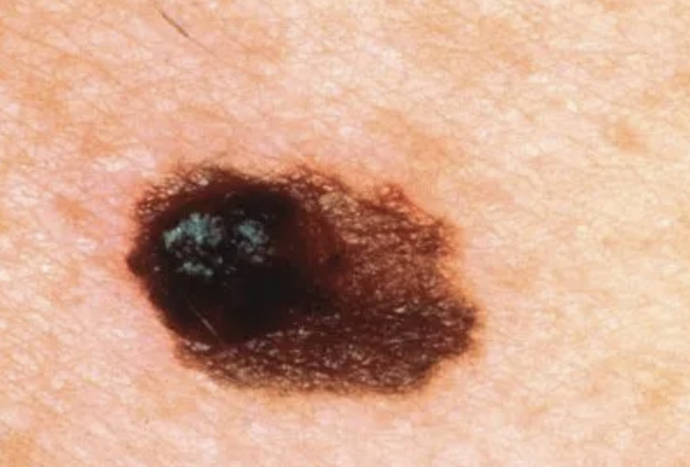
Examples of Malignant Melanomas
C – Colour
Most ordinary moles are a single shade of brown or tan.
One feature that attracts our attention is the presence of several different colours within the same lesion. Shades of dark brown, black, grey, blue, red or white within one mole can sometimes indicate that further assessment is needed.
Equally, colour alone should never be interpreted in isolation. Some perfectly benign moles are naturally very dark, while certain melanomas may appear surprisingly pale.
D – Diameter
Historically, doctors were taught that moles larger than 6 millimetres deserved particular attention.
While this remains a useful guide, modern dermatology has taught us that size alone is not the deciding factor.
Many completely harmless moles are larger than 6 mm, and some early melanomas are significantly smaller.
Rather than focusing solely on the ruler, we now pay much greater attention to whether a lesion is changing over time.
In my opinion, this is the most important letter of all.
Evolution simply means change.
Has the mole changed in size?
Has it changed in colour?
Has it become raised?
Has it started itching?
Has it bled without obvious injury?
Has it developed crusting or failed to heal?
Any genuine change, particularly over a period of weeks or months, deserves professional assessment.
Patients often ask me which feature worries me most. My answer is nearly always the same:
“A mole that is changing.”
Although the ABCDE Guide is an excellent screening tool, experienced clinicians rarely rely on it alone.
When I assess a patient, I also consider:
- Your age.
- Your skin type.
- Your family history.
- Previous sun exposure.
- Whether you’ve experienced blistering sunburns.
- The number of moles you naturally have.
- Whether the lesion looks different from your other moles (the “Ugly Duckling Sign”).
- Dermoscopic features seen under magnification.
- In selected cases, AI-assisted mole analysis.
Every one of these factors helps build a much more complete picture than the ABCDE checklist alone.
What Should You Do If Your Mole Meets One or More ABCDE Criteria?
Don’t panic.
Equally, don’t ignore it.
Most lesions that patients present with after noticing an ABCDE feature prove to be benign. However, because melanoma is highly treatable when detected early, arranging a professional assessment is always the sensible course of action.
It is far better to have a harmless mole examined and be reassured than to spend weeks or months worrying unnecessarily.
Dr Kitchen’s Clinical Pearl
Throughout my career, I have found that patients often remember the ABCDE Rule but forget its purpose.
It was never designed to frighten people into believing every unusual mole is melanoma.
It was designed to encourage people to seek advice when something changes.
Most of the patients I reassure tell me afterwards that the hardest part wasn’t the examination—it was the weeks of uncertainty beforehand.
Knowledge replaces fear, but timely assessment replaces uncertainty.
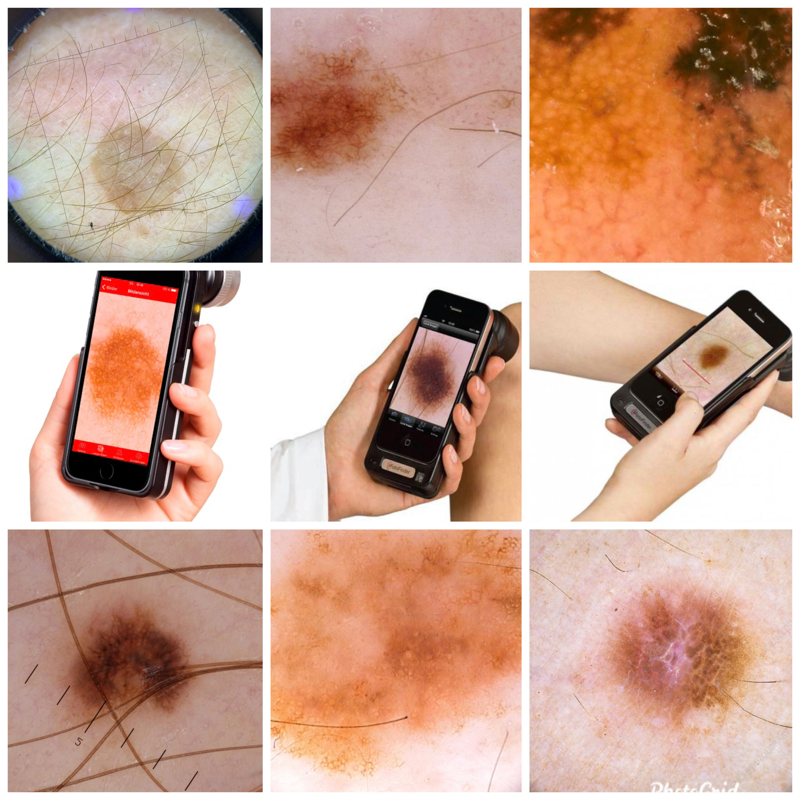
1. A.I. Dermoscopy
3. Mole Mapping Explained
Imagine being able to compare today’s mole with exactly how it appeared one or two years ago. That is the principle behind mole mapping. High-resolution photography, dermoscopy and artificial intelligence now allow us to monitor lesions with remarkable precision. This chapter explains who benefits most, how the technology works, and why digital surveillance has transformed the early detection of melanoma.
Mole Mapping – Seeing Beyond the Naked Eye
Many patients arrive at my clinic believing that examining a mole simply involves “having a quick look.”
In reality, modern skin assessment has advanced enormously over the past two decades.
Today, we can examine skin lesions in far greater detail than ever before, allowing us to identify subtle features that are simply invisible to the naked eye. One of the most valuable advances has been the development of mole mapping and digital dermoscopy.
What Is Mole Mapping?
Mole mapping is a method of carefully documenting and monitoring your moles over time.
Think of it as creating a detailed photographic record of your skin.
Rather than relying on memory, we can compare today’s images with those taken months or even years earlier. Tiny changes that would otherwise go unnoticed can often be detected long before they become obvious to the naked eye.
For many patients, this provides something incredibly valuable—confidence.
Instead of wondering whether a mole has changed, we can compare the images directly.
A dermatoscope is a specialised medical instrument that combines magnification with polarised light, allowing us to look beneath the surface of the skin.
To a patient, a mole may simply appear brown.
Under dermoscopy, however, we can see an intricate world of pigment networks, blood vessels and microscopic structures that help distinguish many benign lesions from those requiring closer attention.
Dermoscopy has transformed the assessment of pigmented skin lesions and is now regarded as an essential part of modern skin cancer diagnosis.
Artificial Intelligence – A Valuable Assistant
In recent years, artificial intelligence (AI) has become another exciting development in dermatology.
Modern AI software can analyse dermoscopic images and compare them with vast databases of previously diagnosed lesions. It is capable of recognising patterns that may not always be immediately obvious and can provide an additional level of analysis.
At our clinic, AI is used as an additional clinical tool, helping to support decision-making.
However, it is important to understand that AI does not replace an experienced clinician.
A computer cannot take a medical history.
It cannot ask when a mole first appeared.
It cannot examine the rest of your skin, appreciate your family history or understand your individual risk factors.
Clinical judgement remains the cornerstone of safe skin assessment.
In my view, the best outcomes are achieved when modern technology is combined with careful clinical examination and many years of experience.
Who Benefits Most from Mole Mapping?
Although anyone can benefit from a detailed skin assessment, mole mapping is particularly valuable for people who:
- Have a large number of moles.
- Have previously had melanoma or another skin cancer.
- Have a strong family history of melanoma.
- Have fair skin that burns easily.
- Have experienced significant sun exposure or repeated childhood sunburn.
- Notice frequent changes in their moles.
- Simply want reassurance and a reliable baseline for future comparison.
For these patients, digital monitoring can provide an additional level of confidence and may help identify important changes at an earlier stage.
How Often Should Mole Mapping Be Performed?
There is no single answer that suits everyone.
The recommended interval depends on your personal level of risk, the number of moles you have and whether any lesions require short-term monitoring.
Some patients only require a single assessment for reassurance.
Others benefit from annual reviews, while a small number of higher-risk individuals may require more frequent monitoring.
Your management plan should always be tailored to your individual circumstances rather than following a one-size-fits-all approach.
The Greatest Benefit Is Often Peace of Mind
One of the most rewarding aspects of mole mapping is not simply detecting abnormalities—it is providing reassurance.
Many patients arrive convinced that a particular mole has changed.
When we compare high-quality images over time, we often discover that it has remained completely stable.
That objective evidence can remove months of unnecessary worry.
Equally, if a lesion has genuinely evolved, we are in a far stronger position to recognise that change early and recommend the most appropriate course of action.
Dr Kitchen’s Clinical Pearl
One lesson I have learnt over nearly four decades is that technology should enhance clinical judgement, never replace it.
Digital dermoscopy and artificial intelligence are remarkable advances in modern medicine, but they are at their most powerful when combined with careful listening, thorough examination and experience gained from assessing thousands of skin lesions.
The most sophisticated camera in the world cannot reassure a patient.
An experienced clinician, supported by the right technology, can often do both.
2. Mole map chart example.
4. Should I Remove My Mole?
One of the most common questions I hear is:
“Doctor, should I just have it removed?”
The answer is not always straightforward.
Some moles should absolutely be removed because they are suspicious or have developed worrying features. Others are removed because they catch on clothing, become irritated during shaving, bleed repeatedly after minor trauma or simply because they have become cosmetically bothersome.
However, many perfectly healthy moles do not need to be removed at all.
One of the most important parts of my consultation is helping patients understand the difference.
When Removal Is Medically Recommended
There are occasions when removal is the safest and most appropriate option.
These include moles that:
- Have changed noticeably in size, shape or colour.
- Display suspicious features on dermoscopic examination.
- Bleed repeatedly without obvious injury.
- Develop persistent crusting or ulceration.
- Cause ongoing symptoms such as itching or pain.
- Cannot be confidently identified as benign during clinical assessment.
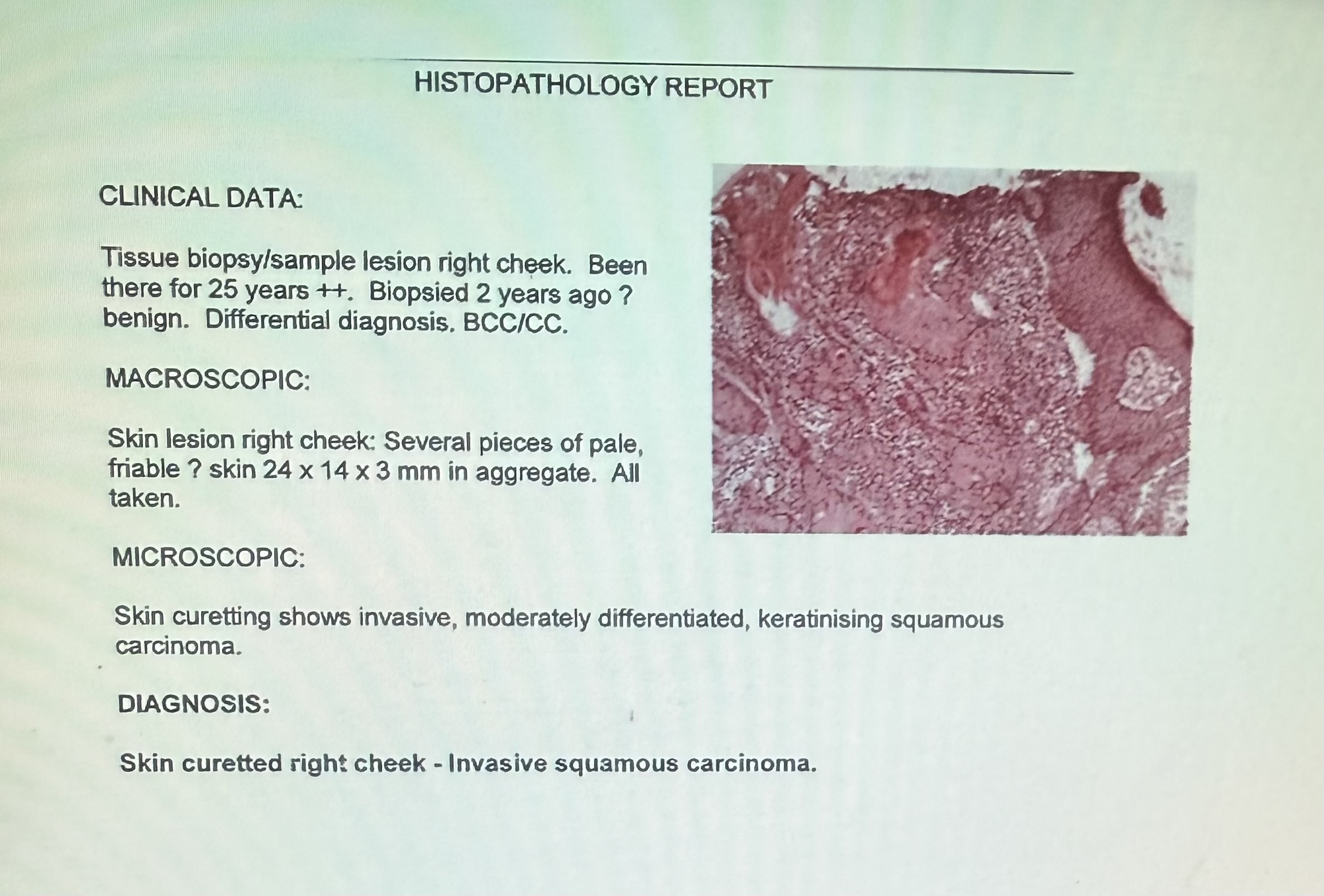
In these situations, removal is recommended because it allows the lesion to be examined properly and, where appropriate, sent for laboratory analysis (histopathology).
For example:-
Early diagnosis remains one of the most important factors in the successful treatment of melanoma and other skin cancers.
When Removal May Be Your Choice
Many patients choose to have a mole removed even though it is entirely benign.
Common reasons include:
- It catches on clothing or jewellery.
- It is repeatedly cut while shaving.
- It rubs against bra straps or waistbands.
- It becomes inflamed after minor trauma.
- It affects confidence because of its appearance.
- It has become increasingly prominent with age.
These are all perfectly reasonable reasons to consider removal.
Cosmetic concerns should never be dismissed. If a lesion is affecting your confidence or quality of life, that matters.
When I May Recommend Leaving It Alone
Not every mole benefits from removal.
If a mole has remained unchanged for many years, shows completely benign features and causes no symptoms, there may be little medical advantage in removing it.
Every procedure, however small, carries the possibility of leaving a scar.
For that reason, I always balance the benefits of removal against the likely cosmetic outcome.
Sometimes the best advice I can give is:
“This mole is completely harmless and I don’t believe removing it will improve matters.”
Patients are often surprised by this advice, but good medicine is about recommending what is genuinely in your best interests rather than simply performing a procedure because it has been requested.
Will Mole Removal Leave a Scar?
This is probably the second most common question I am asked.
The honest answer is:
Any procedure that removes skin has the potential to leave some degree of scarring.
Fortunately, modern surgical and radiofrequency techniques usually produce scars that fade remarkably well over time, particularly when the procedure is carefully planned and appropriate aftercare advice is followed.
Many scars become difficult to notice after several months.
The final appearance depends on many factors, including:
- The size and depth of the lesion.
- Its location on the body.
- Your natural healing response.
- Whether the wound is under tension.
- How well the aftercare instructions are followed.
Part of my role is to discuss realistic expectations before any treatment takes place.
Will the Mole Grow Back?
Most moles that are completely removed do not return.
Occasionally, if a small number of pigment cells remain within the skin, some pigmentation may reappear over time.
This does not automatically indicate skin cancer. It simply reflects the fact that a small number of melanocytes remain in the treated area.
If any recurrent pigmentation develops, it should always be reviewed so that it can be assessed appropriately.
Should Every Mole Be Sent to the Laboratory?
Not necessarily.
This is a question that often causes confusion.
When a mole appears clinically suspicious, or there is uncertainty about the diagnosis, laboratory examination under the microscope (histopathology) is strongly recommended.
However, many lesions removed for cosmetic reasons have entirely typical benign features following careful clinical assessment and dermoscopy.
The decision to send tissue for laboratory analysis should always be based on good clinical judgement and current practice, taking into account the appearance of the lesion and the reason for removal.
I will always discuss this with you before treatment so that you understand the rationale behind the recommendation.
Making the Right Decision
Removing a mole should never be an automatic decision.
It should be an informed decision.
That means understanding:
- Why the mole is being removed.
- Whether removal is medically necessary or elective.
- The likely cosmetic result.
- The small risks associated with any procedure.
- Whether laboratory analysis is advisable.
Only then can you decide what is right for you.
Dr Kitchen’s Clinical Pearl
One of the greatest misconceptions is that every mole should simply be removed “just in case.”
After examining many thousands of skin lesions, I have learnt that careful assessment almost always comes before treatment.
Sometimes the best outcome is removal.
Sometimes the best outcome is reassurance.
Knowing the difference is one of the most valuable parts of an experienced clinical assessment.
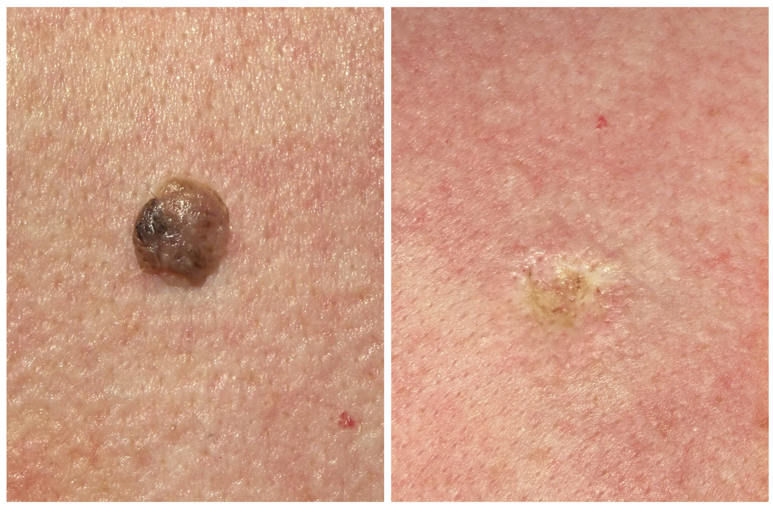
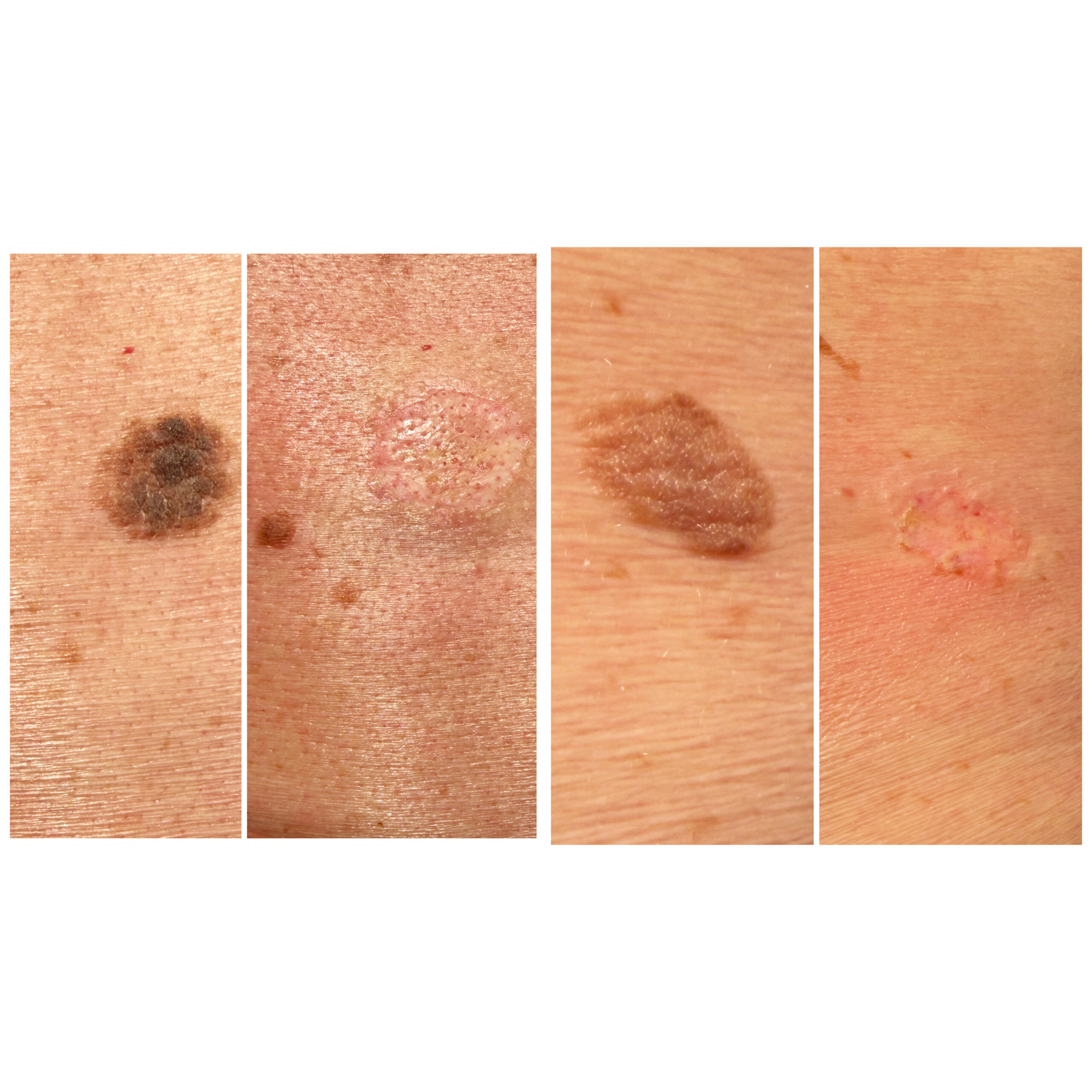
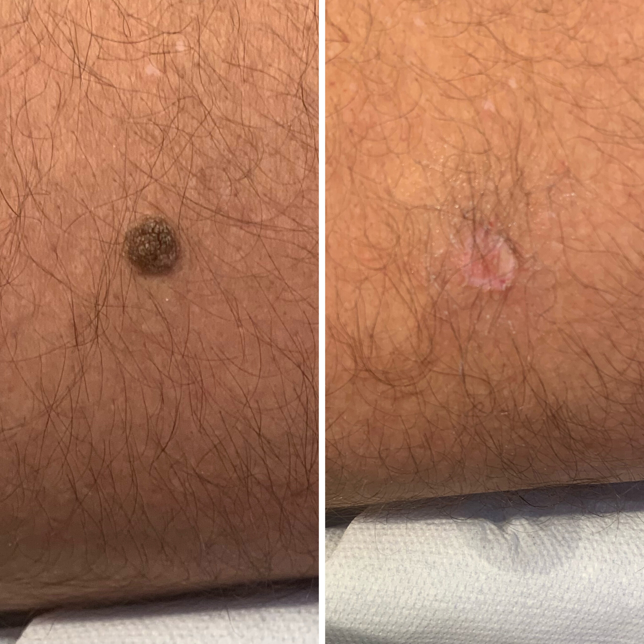
3. Mole removal using 4. Classic Seb’ keratosis immediately
Radio-Frequency loop before & after
If you’d like to understand how we use digital photography, dermoscopy and A.I. to monitor changing moles, read my guide on Mole Mapping Explained.
5. Skin Cancer Prevention
The best treatment for skin cancer is to prevent it developing in the first place. Yet prevention is often misunderstood. Contrary to popular belief, avoiding the sun altogether is neither practical nor necessarily desirable. Sensible sun exposure, appropriate protection, avoiding burning and recognising individual risk factors form the cornerstone of healthy skin. Prevention is about balance, not fear.
Skin Cancer Prevention – Protecting Your Skin Without Living in Fear of the Sun
When people think about skin cancer prevention, they often imagine one simple message:
“Stay out of the sun.”
In reality, the picture is far more balanced than that.
Sunlight is not the enemy.
It plays an important role in our overall health, contributing to vitamin D production, supporting our natural body clock and positively influencing mood and wellbeing.
The problem is not sensible sun exposure.
The problem is too much ultraviolet (UV) radiation, particularly when it results in repeated sunburn.
Like many aspects of medicine, skin health is about achieving the right balance rather than living at either extreme.
Understanding Ultraviolet (UV) Radiation
The sun emits several types of ultraviolet radiation, but the two most relevant to skin health are UVA and UVB.
Both penetrate the skin and contribute to premature ageing and DNA damage, although in different ways.
Over many years, repeated UV exposure can damage the genetic material within skin cells. Fortunately, our bodies possess remarkable repair mechanisms that correct much of this damage every day.
Occasionally, however, damaged cells escape these repair systems.
It is this accumulation of genetic damage over many years that increases the risk of developing skin cancers.
Importantly, this process usually occurs gradually over decades rather than after a single afternoon in the sunshine.
Why Childhood Sunburn Matters
One of the strongest risk factors for melanoma is a history of repeated blistering sunburn, particularly during childhood and adolescence.
Young skin is especially vulnerable to ultraviolet damage.
For this reason, protecting children from burning remains one of the most important investments parents can make in their long-term skin health.
That does not mean children should never play outside.
It simply means avoiding prolonged exposure when the sun is strongest and preventing sunburn whenever possible.
Who Is Most at Risk?
Although anyone can develop skin cancer, certain people have a higher lifetime risk.
These include individuals who:
- Have fair skin that burns easily.
- Have blue or green eyes.
- Have naturally blonde or red hair.
- Have a large number of moles.
- Have a personal or family history of melanoma.
- Have experienced repeated severe sunburn.
- Spend long periods working or exercising outdoors.
- Have a weakened immune system.
If several of these factors apply to you, regular skin examinations become even more important.
Practical Ways to Protect Your Skin
Fortunately, reducing your risk of skin cancer does not require avoiding outdoor life altogether.
Simple measures include:
- Avoiding sunburn whenever possible.
- Seeking shade during the hottest part of the day if spending prolonged periods outdoors.
- Wearing a wide-brimmed hat and UV-protective clothing when appropriate.
- Using a broad-spectrum sunscreen on exposed skin during prolonged outdoor activities.
- Reapplying sunscreen regularly, particularly after swimming or heavy perspiration.
- Taking extra care near water, snow and sand, where UV radiation is reflected.
Protection should be practical rather than obsessive.
The aim is to enjoy outdoor life safely.
Sunbeds – A Risk Not Worth Taking
Unlike natural sunlight, there is no health benefit to using commercial tanning beds.
Numerous studies have shown that sunbed use increases the risk of melanoma and other skin cancers, particularly when started at a young age.
For this reason, I strongly advise against their use.
A tan should never be regarded as a sign of healthy skin.
It is simply evidence that the skin has responded to ultraviolet exposure.
Get to Know Your Own Skin
One of the most effective forms of skin cancer prevention costs nothing.
Become familiar with your own skin.
Spend a few minutes every month or two looking for any new lesions or changes in existing moles.
Remember the ABCDE Guide and the Ugly Duckling Sign discussed earlier in this Knowledge Hub.
Most changes prove entirely harmless.
Occasionally, however, recognising a suspicious lesion early can make an enormous difference.
Early Detection Saves Lives
Modern treatments for melanoma have improved dramatically.
When detected early, melanoma is often highly treatable.
This is why I encourage patients not to ignore persistent changes, but equally not to panic.
Concern should prompt assessment.
Not fear.
Dr Kitchen’s Clinical Pearl
One of the greatest misconceptions I encounter is that skin cancer prevention means avoiding the sun completely.
I don’t believe that is either realistic or desirable.
After many years in clinical practice, my advice remains remarkably simple:
Enjoy the outdoors. Respect the sun. Avoid burning. Know your own skin.
Most importantly, if something changes or simply doesn’t look quite right, have it assessed.
Knowledge, common sense and early diagnosis remain our most powerful forms of protection.
5. Beware and protect
6. Seborrhoeic Keratoses
Often described as “barnacles of ageing,” seborrhoeic keratoses are among the commonest benign skin growths I encounter. They can appear alarming, suddenly increase in number and are frequently mistaken for skin cancer. Fortunately, they are entirely benign. We’ll explain why they occur, when they should be assessed and the safest ways to remove them with excellent cosmetic outcomes.
Seborrhoeic Keratoses – The Great Mimic
One of the commonest reasons patients attend my clinic is because they have discovered what they believe to be a new mole or, understandably, fear they may have developed skin cancer.
Very often, the diagnosis is something far less worrying.
The lesion turns out to be a seborrhoeic keratosis.
Although the name sounds rather alarming, seborrhoeic keratoses are completely benign skin growths and are among the commonest skin lesions I diagnose.
In fact, most people will develop at least one during their lifetime, and many will develop dozens as they get older.
What Is a Seborrhoeic Keratosis?
A seborrhoeic keratosis (often abbreviated to SK) is a harmless overgrowth of the outer layer of the skin.
Unlike moles, they do not arise from pigment-producing cells (melanocytes).
Instead, they develop from ordinary skin cells called keratinocytes, which gradually build up to form a slightly raised plaque on the surface of the skin.
They are entirely benign.
Importantly, they are not skin cancer and they do not turn into skin cancer.
Why Do They Develop?
The exact cause remains unknown, but several factors appear to contribute.
Age is by far the strongest influence.
For this reason they are sometimes affectionately referred to as the “barnacles of ageing.”
While that description may raise a smile, it reflects the fact that these lesions tend to appear gradually over many years.
They also have a strong genetic tendency.
If your parents developed numerous seborrhoeic keratoses, there is a good chance that you will too.
Sun exposure may play a contributory role in some patients, although many develop in areas that receive very little sunlight.
What Do They Look Like?
Seborrhoeic keratoses vary enormously in appearance.
They may be:
- Light tan, brown or almost black.
- Smooth or rough.
- Flat or raised.
- Small pinhead lesions or several centimetres across.
- Single or present in large numbers.
One of their most recognisable features is their appearance.
Many dermatologists describe them as looking as though they have been “stuck onto the skin.”
Some have a waxy appearance.
Others resemble a drop of candle wax.
Older lesions may become rough, crumbly or wart-like.
Because they can become very dark in colour, they are frequently mistaken for melanoma by patients.
Where Do They Occur?
Seborrhoeic keratoses most commonly develop on:
- The chest.
- The back.
- The shoulders.
- The face.
- The scalp.
- The neck.
They rarely occur on the palms of the hands or the soles of the feet.
Many patients notice that they seem to appear “overnight.”
In reality, they usually develop very gradually until one day they simply catch your attention.
Can They Cause Symptoms?
Although entirely benign, seborrhoeic keratoses can become troublesome.
Some itch.
Some catch on clothing.
Some become inflamed after rubbing against bra straps or collars.
Occasionally they become sore or bleed after accidental trauma.
These symptoms do not necessarily mean they are dangerous.
More often, they simply indicate irritation.
How Are They Diagnosed?
In many cases, an experienced clinician can recognise a seborrhoeic keratosis during routine examination.
However, because some pigmented lesions can closely resemble melanoma, careful assessment is essential.
I routinely examine suspicious lesions using dermoscopy, which allows me to see microscopic structures beneath the surface of the skin that are invisible to the naked eye.
Where appropriate, digital imaging and AI-assisted analysis may also provide additional information to support the assessment.
If there is ever uncertainty about the diagnosis, further investigation or removal may be recommended.
Should They Be Removed?
Because seborrhoeic keratoses are harmless, removal is not medically necessary.
However, many patients choose treatment because the lesion:
- Catches on clothing.
- Becomes repeatedly irritated.
- Is cosmetically noticeable.
- Continues to enlarge.
- Causes anxiety.
Modern radiofrequency surgery usually allows these lesions to be removed quickly, often with an excellent cosmetic outcome.
The most appropriate treatment depends on the size, thickness and location of the lesion.
This is something we discuss during your consultation.
Can They Come Back?
Once an individual seborrhoeic keratosis has been completely removed, it usually does not return.
However, because the tendency to develop them is often inherited, it is perfectly possible for new seborrhoeic keratoses to appear elsewhere over time.
This does not indicate that treatment has failed.
It simply reflects your natural skin type.
Dr Kitchen’s Clinical Pearl
If I had to choose the single lesion that causes the greatest unnecessary anxiety, it would probably be the seborrhoeic keratosis.
Every week I meet patients convinced they have developed skin cancer, only to discover that the lesion is completely benign.
That moment—when I can reassure someone that what they feared is not what they have—is one of the most rewarding parts of my work.
It also reminds us why self-diagnosis has its limits.
Modern technology, careful examination and clinical experience together provide far more reliable answers than internet searches ever can.
6. Seborrhoeic wart on scalp 7. Examples of before & after RF ablation for Seb K’s. Often have a waxy appearance and well defined border… “a stuck on appearance”
8. Seborrhoeic Wart on leg
Classic Seb’ K’s before and immediately after Radio-Frequency Ablation
7. Skin Tags _ Harmless but Annoying
Tiny though they are, skin tags can cause disproportionate annoyance. Whether they rub against clothing, catch on jewellery or simply affect confidence, patients often ask whether they can safely be removed. This chapter discusses why skin tags develop, why they are associated with friction and sometimes insulin resistance, and how modern radio-frequency techniques usually remove them quickly with minimal discomfort.
Skin Tags – Small Lesions That Can Become a Big Irritation
Skin tags are among the commonest benign skin growths I see in clinical practice.
Although they are completely harmless, they can become surprisingly troublesome. Some catch on clothing, others rub against jewellery, become irritated during shaving or simply affect confidence because of where they are located.
The good news is that skin tags are not skin cancer, they do not become cancerous, and in most cases they can be removed quickly and safely if they are causing problems.
What Is a Skin Tag?
A skin tag, known medically as an acrochordon, is a small, soft growth that projects from the surface of the skin.
They are usually attached by a narrow stalk, giving them their characteristic “dangling” appearance.
Most skin tags measure only a few millimetres across, although some gradually enlarge over time and may become more noticeable.
They are composed of completely normal skin tissue and are entirely benign.
Why Do Skin Tags Develop?
The exact cause is not fully understood, but several factors appear to contribute.
The most important is friction.
Skin tags commonly develop where skin repeatedly rubs against skin or clothing. This explains why they are so frequently found in natural body folds.
Other recognised factors include:
- Increasing age.
- A family tendency.
- Being overweight.
- Pregnancy, when hormonal changes may encourage new skin tags to develop.
- Conditions associated with insulin resistance, such as type 2 diabetes.
Having a skin tag does not mean you have diabetes, but people with multiple skin tags may benefit from discussing their general health with their GP, particularly if they have other risk factors.
Where Do Skin Tags Usually Occur?
Skin tags are most commonly found:
- On the neck.
- In the armpits.
- Around the eyelids.
- Beneath the breasts.
- In the groin.
- Along areas where clothing causes repeated friction.
Although they can occur almost anywhere, these areas account for the vast majority of cases I see.
Are Skin Tags Dangerous?
The simple answer is no.
Skin tags are completely benign and do not develop into skin cancer.
They usually remain unchanged for many years, although they may slowly enlarge over time.
Occasionally, a skin tag twists on its narrow stalk, temporarily cutting off its own blood supply. When this happens it may become painful, swollen, dark in colour or eventually fall off naturally.
Although this can look alarming, it is rarely a cause for concern.
Should I Remove My Skin Tag?
Not necessarily.
If a skin tag is small, causes no symptoms and does not bother you cosmetically, there is no medical need to remove it.
However, many people choose treatment because the skin tag:
- Catches on clothing or jewellery.
- Bleeds after shaving.
- Becomes irritated repeatedly.
- Is unsightly.
- Makes them self-conscious.
These are all perfectly reasonable reasons to consider removal.
How Are Skin Tags Removed?
Several treatment methods are available, depending on the size and location of the skin tag.
In my practice, one of the most effective techniques is high-frequency radiofrequency surgery, which allows the skin tag to be removed with remarkable precision while minimising damage to the surrounding skin.
The procedure is usually very quick.
Most patients are surprised by how little discomfort they experience, and healing is generally straightforward with appropriate aftercare.
The most suitable treatment is always discussed during your consultation.
Can I Remove a Skin Tag Myself?
Many over-the-counter products and internet remedies claim to remove skin tags.
While some may work in selected cases, I would always advise caution.
Not every lump is a skin tag.
Occasionally, other benign skin lesions—or, much less commonly, skin cancers—can resemble skin tags.
For this reason, obtaining the correct diagnosis before attempting treatment is always the safest approach.
Will They Come Back?
Once a skin tag has been completely removed, it usually does not return.
However, people who are naturally prone to developing skin tags may continue to form new ones elsewhere over time.
This is simply part of the way their skin behaves and does not indicate that treatment has failed.
Dr Kitchen’s Clinical Pearl
Skin tags may be tiny, but they often cause a surprising amount of frustration.
One of the nicest moments in clinic is removing a lesion that has caught on a necklace or shirt collar for years and hearing the patient say, “I wish I’d had that done ages ago.”
Sometimes the smallest procedures make the biggest difference to everyday comfort and confidence.
As with all skin lesions, the first step is making the correct diagnosis.
Once we know exactly what we’re dealing with, the most appropriate treatment usually becomes very straightforward.
8. Skin Tag removal….so common but gone in minutes

8. Cysts
A lump beneath the skin often causes understandable concern. In reality, many prove to be epidermoid cysts—small sacs containing keratin that have become trapped beneath the skin’s surface. They are usually harmless but may become inflamed, infected or cosmetically prominent. Here we discuss diagnosis, common misconceptions and why squeezing them at home is rarely a good idea.
Skin Cysts – Understanding the Lump Beneath the Skin
Discovering a lump beneath your skin can be worrying.
For many people, the first thought is understandably, “Could it be something serious?”
Fortunately, one of the commonest lumps I see in clinical practice is a skin cyst, and in the overwhelming majority of cases these are completely benign.
Although they may gradually enlarge, become inflamed or occasionally become infected, they are not skin cancer and rarely represent a serious medical problem.
The key is making the correct diagnosis.
What Is a Skin Cyst?
A skin cyst is a small sac that develops beneath the surface of the skin.
Most contain keratin—a soft, cheese-like material made from the same protein found in our skin, hair and nails.
Many people refer to these as sebaceous cysts, but this term is not entirely accurate.
Most are actually epidermoid cysts (sometimes called epidermal inclusion cysts), arising from cells within the outer layer of the skin rather than from the sebaceous (oil) glands themselves.
Although the terminology sounds complicated, the important message is simple:
They are very common.
They are usually harmless.
Why Do Cysts Develop?
Skin cells are continually produced within the outer layer of the skin.
Occasionally, instead of shedding normally, some of these cells become trapped beneath the surface.
They continue producing keratin, which gradually accumulates inside a small sac, causing the cyst to enlarge over time.
Sometimes a blocked hair follicle or previous minor injury to the skin may contribute, although many cysts develop for no obvious reason.
They are not caused by poor hygiene.
Nor are they contagious.
What Does a Cyst Feel Like?
Most cysts have several characteristic features.
They are usually:
- Round or oval.
- Smooth.
- Soft to firm in consistency.
- Freely movable beneath the skin.
- Slow growing.
- Usually painless unless inflamed.
Many also have a tiny central opening, known as a punctum, which represents the original blocked skin pore.
This small feature often helps distinguish a cyst from other types of skin lump.
Where Do Cysts Occur?
Cysts can develop almost anywhere on the body but are particularly common on:
- The scalp.
- The face.
- The neck.
- The shoulders.
- The upper back.
- The chest.
Some remain only a few millimetres in size, while others slowly enlarge over several years.
Why Do Cysts Sometimes Become Painful?
Although a cyst itself is harmless, problems may arise if it becomes inflamed or infected.
The cyst may suddenly become:
- Red.
- Tender.
- Swollen.
- Warm to the touch.
Occasionally it may discharge thick, unpleasant-smelling keratin or pus if it ruptures through the skin.
This can be alarming but is usually manageable with appropriate treatment.
Attempting to squeeze or burst a cyst yourself often makes matters worse and may increase the risk of infection or scarring.
Should Every Cyst Be Removed?
Not necessarily.
Many cysts remain small, painless and unchanged for years.
If they are causing no symptoms and the diagnosis is certain, simple observation is often perfectly appropriate.
However, removal may be recommended if the cyst:
- Continues to enlarge.
- Becomes repeatedly inflamed.
- Causes discomfort.
- Is cosmetically troublesome.
- Frequently catches during shaving or brushing.
- Creates uncertainty about the diagnosis.
Complete removal of the cyst wall is important if recurrence is to be minimised.
How Are Cysts Removed?
The most appropriate technique depends on the size and location of the cyst.
Smaller cysts may often be removed under local anaesthetic during a straightforward outpatient procedure.
Larger cysts, particularly those that have been repeatedly inflamed, may require a slightly more involved surgical approach.
The aim is always to remove the cyst completely while achieving the best possible cosmetic outcome.
Can a Cyst Come Back?
If the entire cyst wall is successfully removed, recurrence is uncommon.
However, if part of the lining remains beneath the skin, the cyst may gradually reform.
This is one reason why repeated squeezing or partial drainage is rarely a long-term solution.
When Should I Seek Medical Advice?
Although most cysts are harmless, you should arrange an assessment if:
- The lump is growing rapidly.
- It becomes persistently painful.
- It repeatedly becomes infected.
- The diagnosis is uncertain.
- It interferes with your daily activities.
- You are simply worried about what it might be.
Remember, not every lump beneath the skin is a cyst.
Obtaining the correct diagnosis is always the first step towards choosing the most appropriate treatment.
Dr Kitchen’s Clinical Pearl
One of the commonest things I hear in clinic is, “I’ve had this lump for years, but it’s suddenly become sore.”
In most cases, this doesn’t mean it has become dangerous.
More often, it simply means the cyst has become inflamed or irritated.
The important lesson is this:
Never ignore a changing lump, but equally don’t assume the worst.
A careful examination usually provides the answer—and very often, the reassurance that patients have been hoping for.
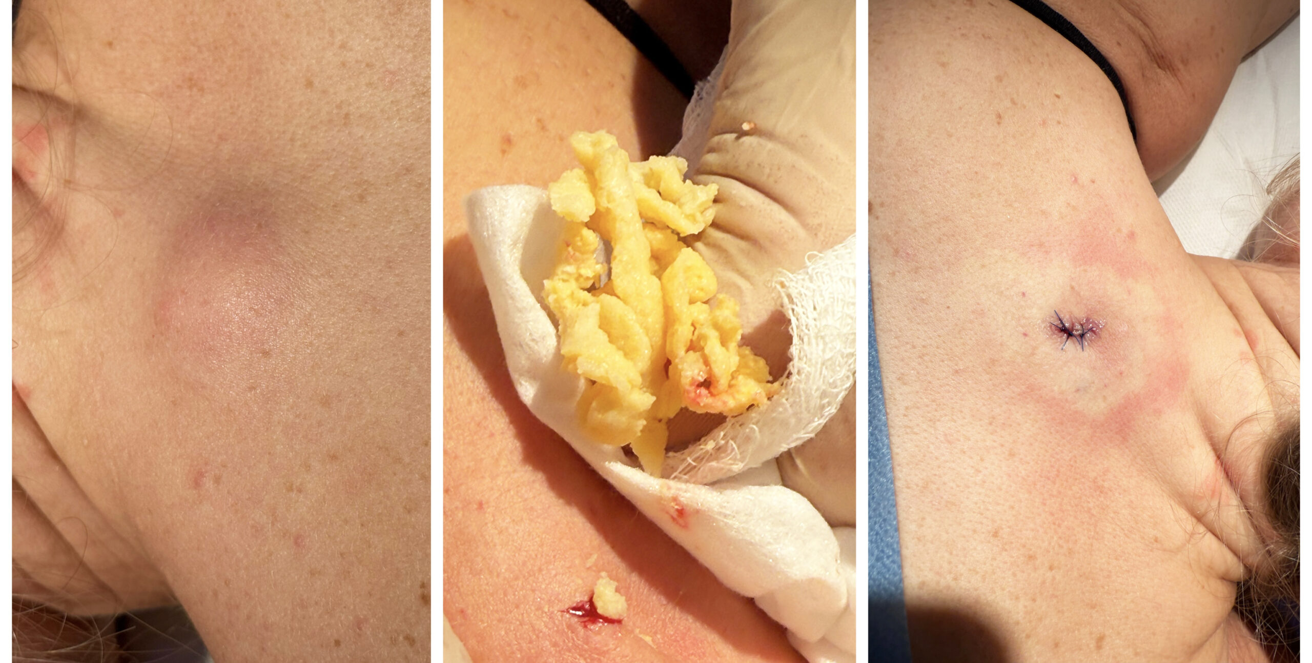
9. Sebaceous Cysts drainage. So vital to get the capsule out.
10. Classic Sebaceous cyst drainage
9. Lipomas
Soft, mobile fatty lumps known as lipomas are extremely common and almost always benign. Nevertheless, any new lump deserves careful assessment to ensure the diagnosis is correct. We’ll explore how lipomas differ from more concerning growths, when imaging is appropriate, and the circumstances in which surgical removal offers both reassurance and symptom relief.
Lipomas – The Common “Fatty Lump” Beneath the Skin
Few things cause more anxiety than discovering a lump beneath your skin.
If you’re unsure whether a lump is a lipoma or a cyst, read my guide to
One of the first questions patients ask is:
“Could it be cancer?”
Fortunately, one of the commonest soft tissue lumps I diagnose is a lipoma, and the overwhelming majority are entirely benign.
Lipomas are extremely common, often develop slowly over many years and, in most cases, never cause any serious health problems.
The important step is making an accurate diagnosis.
What Is a Lipoma?
A lipoma is a benign growth of normal fat cells.
Rather than spreading throughout the fatty tissue beneath the skin, a small group of fat cells slowly gathers together to form a soft, rounded lump.
Lipomas are not skin cancer.
They are not infectious.
They do not spread from one part of the body to another.
In most cases, they remain harmless throughout life.
Why Do Lipomas Develop?
The exact reason why lipomas develop remains uncertain.
We do know that they are often inherited, and it is not uncommon for several members of the same family to develop them.
Contrary to popular belief, lipomas are not caused by being overweight.
Slim people develop lipomas just as commonly as those carrying excess weight.
Occasionally, patients notice a lipoma developing after a minor injury, although whether trauma actually causes the lump or simply draws attention to one that was already present remains uncertain.
What Does a Lipoma Feel Like?
Lipomas have several characteristic features.
They are usually:
- Soft and slightly rubbery.
- Smooth.
- Rounded or oval.
- Easily moved beneath the skin with gentle pressure.
- Painless.
- Slow growing.
Many patients describe them as feeling like a small rubber ball beneath the skin.
Their soft, mobile nature often helps distinguish them from other types of lump.
Where Do Lipomas Occur?
Lipomas can develop almost anywhere that fat exists beneath the skin.
They are particularly common on:
- The shoulders.
- The upper back.
- The neck.
- The upper arms.
- The thighs.
- The trunk.
Some people develop a single lipoma, while others naturally form several over their lifetime.
Do Lipomas Become Cancerous?
This is probably the commonest question I am asked.
The reassuring answer is that ordinary lipomas do not transform into cancer.
However, not every lump beneath the skin is a lipoma.
This is why any new lump, rapidly enlarging lump or unusually firm lump deserves proper assessment.
Very occasionally, further investigation such as ultrasound imaging or referral to a specialist may be recommended if the diagnosis is uncertain.
Fortunately, these situations are uncommon.
Should Every Lipoma Be Removed?
No.
Many lipomas remain unchanged for years and never require treatment.
Removal is usually considered if the lipoma:
- Continues to enlarge.
- Causes discomfort.
- Presses against nearby structures.
- Restricts movement.
- Is cosmetically noticeable.
- Causes anxiety because of its appearance.
Some patients simply find the lump irritating or dislike the appearance beneath clothing.
These are entirely reasonable reasons to discuss treatment.
How Are Lipomas Removed?
Most lipomas can be removed under local anaesthetic as a straightforward outpatient procedure.
The exact technique depends on the size and position of the lump.
The aim is to remove the lipoma completely while achieving the best possible cosmetic result.
Following removal, the lump may be sent for laboratory examination where clinically appropriate, particularly if there is any uncertainty regarding the diagnosis.
Will It Come Back?
If a lipoma has been completely removed, recurrence is uncommon.
However, because some people have an inherited tendency to develop lipomas, new ones may appear elsewhere over time.
This does not mean the original treatment has failed.
It simply reflects the way your body naturally forms these benign fatty growths.
When Should I Arrange an Assessment?
Although lipomas are usually harmless, you should seek medical advice if:
- You notice a new lump beneath the skin.
- A lump is growing rapidly.
- It becomes painful.
- It feels unusually firm or fixed.
- It lies deep within the tissues.
- You are simply unsure what it is.
Most lumps prove to be benign.
Nevertheless, obtaining the correct diagnosis is always the safest and most reassuring approach.
Dr Kitchen’s Clinical Pearl
Over the years, I have learnt that patients often fear the unknown far more than the diagnosis itself.
Many arrive convinced the lump beneath their skin represents something serious.
Very often, I am able to reassure them that it is simply a harmless lipoma.
Those moments are a reminder that reassurance is one of the most valuable treatments a doctor can provide—but reassurance should always be based upon careful examination and an accurate diagnosis.
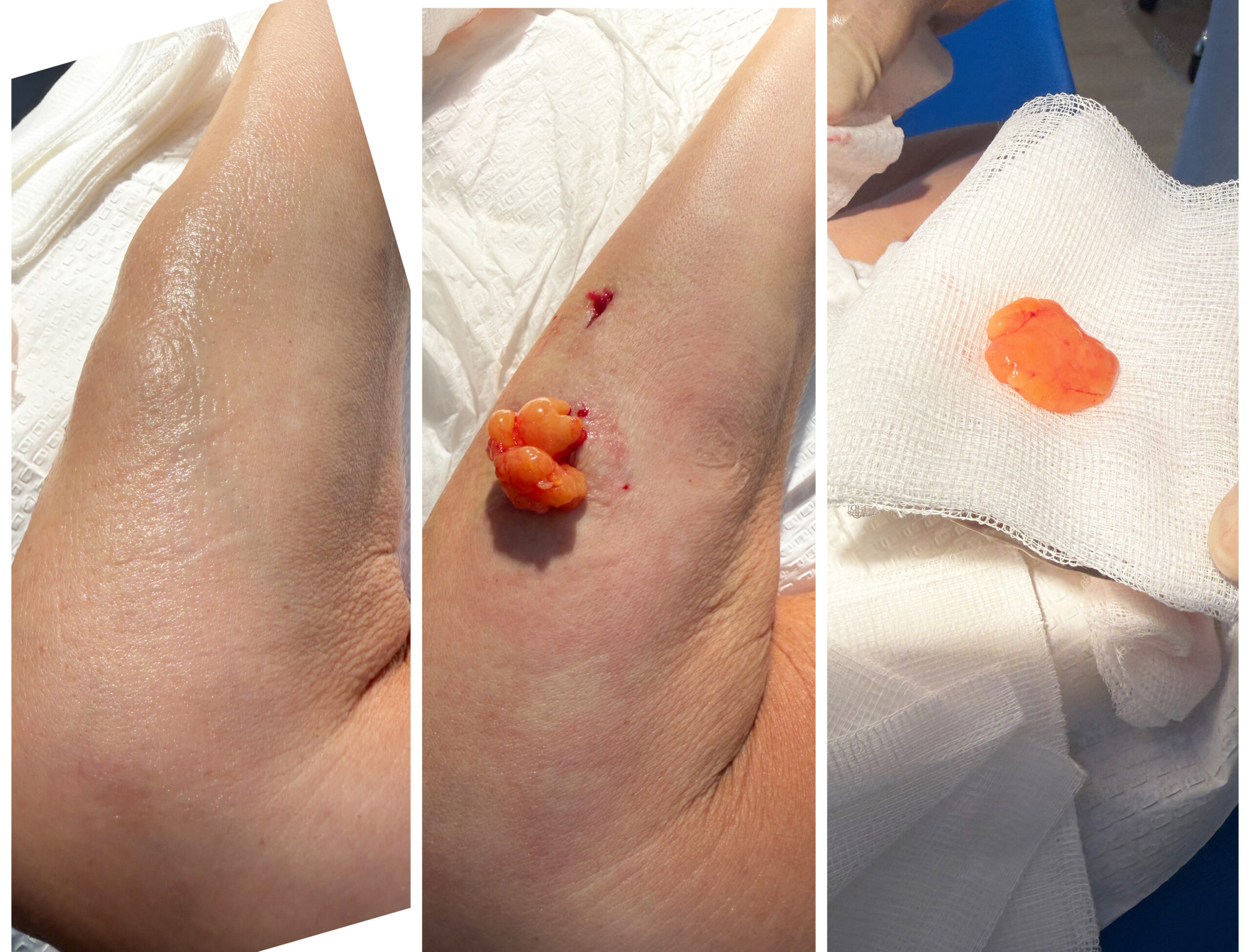
11. Multiple Lipomas are very common but easily removed.
12. Lipomas are very common on the limbs; here a sizeable lesion on the forearm.
10. Warts & Verrucae
Caused by the human papillomavirus, warts and verrucae are surprisingly resilient. Many eventually disappear without treatment, while others persist for years despite over-the-counter remedies. Understanding why the immune system sometimes clears them and sometimes doesn’t helps explain why no single treatment is universally successful. We’ll review the evidence behind cryotherapy, radio-frequency surgery, topical therapies and simple measures to reduce spread.
Warts & Verrucae – Common, Contagious and Usually Harmless
Few skin conditions cause as much frustration as warts and verrucae.
Although they are completely benign, they can persist for months or even years, often resisting a wide variety of over-the-counter treatments. It is not unusual for patients to tell me they have tried “everything” without success.
The good news is that warts and verrucae are not skin cancer, they do not become cancerous, and there are a number of effective treatment options available when they become troublesome.
The first step, however, is understanding exactly what they are.
What Are Warts and Verrucae?
Warts are small benign skin growths caused by infection with the Human Papillomavirus (HPV).
The virus infects the outer layer of the skin, causing the cells to multiply more rapidly than normal and producing the familiar rough, raised appearance.
A verruca is simply a wart that develops on the sole of the foot.
Although the name sounds different, the underlying cause is exactly the same.
How Do You Catch Them?
The virus spreads through direct skin contact or indirectly from contaminated surfaces such as communal changing rooms, swimming pools and shower areas.
Fortunately, most people are naturally resistant to infection, and simply coming into contact with the virus does not guarantee that a wart will develop.
Small breaks in the skin make infection more likely.
This explains why verrucae are particularly common in children, teenagers, swimmers and those who regularly walk barefoot in communal areas.
What Do They Look Like?
Warts can vary considerably in appearance.
They may be:
- Small, rough, raised bumps.
- Flesh coloured, white or slightly brown.
- Single or present in clusters.
- Smooth on the face but rough elsewhere.
Verrucae often look slightly different because pressure from standing pushes them into the sole of the foot.
They may appear flat with a rough central surface and tiny black dots, which are actually small blood vessels rather than dirt.
Some become painful when walking because body weight presses them into the deeper tissues.
Will They Go Away Naturally?
Yes.
One of the most interesting things about warts is that they often disappear without any treatment at all.
Your immune system gradually recognises the virus and eventually eliminates it.
Unfortunately, this process can be very slow.
Some disappear within months.
Others remain for several years.
The challenge is that no one can accurately predict when an individual wart will resolve naturally.
Should Every Wart Be Treated?
Not necessarily.
If a wart is painless, not spreading and causing no cosmetic concern, simply leaving it alone is often a perfectly reasonable option.
Treatment is usually considered if:
- The wart is painful.
- It interferes with walking or sport.
- It is spreading.
- It catches during shaving.
- It is cosmetically embarrassing.
- It has persisted despite self-treatment.
Treatment Options
Many over-the-counter preparations contain salicylic acid, which gradually softens and removes infected skin.
When used consistently over several weeks or months, these products can be very effective.
Other treatment options include:
- Cryotherapy (freezing).
- Radiofrequency surgery.
- Curettage in selected cases.
- Other specialist treatments where appropriate.
No single treatment works for every wart because the success of treatment depends partly on your immune system recognising and clearing the virus.
This is why patience is often just as important as the treatment itself.
Can Warts Be Prevented?
Although it is impossible to prevent every wart, a few simple measures may reduce the risk.
These include:
- Wearing footwear in communal showers and swimming pools.
- Avoiding picking at existing warts.
- Keeping feet clean and dry.
- Avoiding sharing towels, pumice stones or nail equipment.
- Covering warts during swimming where appropriate.
When Should I Seek Medical Advice?
Most warts are straightforward to recognise.
However, you should arrange an assessment if:
- The diagnosis is uncertain.
- The lesion changes rapidly.
- It bleeds repeatedly.
- It becomes persistently painful.
- It fails to respond to treatment.
- You have diabetes or reduced circulation and develop a painful foot lesion.
Occasionally, skin cancers and other lesions can resemble warts.
Obtaining the correct diagnosis is therefore the first and most important step before treatment begins.
Dr Kitchen’s Clinical Pearl
Patients often ask me:
“Doctor, what’s the best treatment for warts?”
My answer is always the same.
The best treatment depends not only on the wart itself, but also on the individual sitting in front of me.
Age, general health, previous treatments, the location of the wart and how troublesome it has become all influence the decision.
After many years in practice, I have learnt that there is rarely a single “magic” treatment.
Good results come from choosing the right treatment for the right patient at the right time.
Sometimes the most effective prescription is patience.
Sometimes it is intervention.
Knowing the difference is where experience matters.
13. Warts (Viral origin) very common on hands and feet
14. Annoying wart inside nasal meatus.
15. Classic wart on hands can be visually disfiguring. Easily removed with Radio-frequency ablation
16. Plantar warts (Verruccae) Unless caught early most over-the-counter treatments DO NOT WORK ; Chronically they can form nests under the weight bearing skin of the feet and after ablation may take up to 6 weeks to heal over.
11. Xanthelasma
Xanthelasma – Understanding Yellow Cholesterol Deposits Around the Eyes
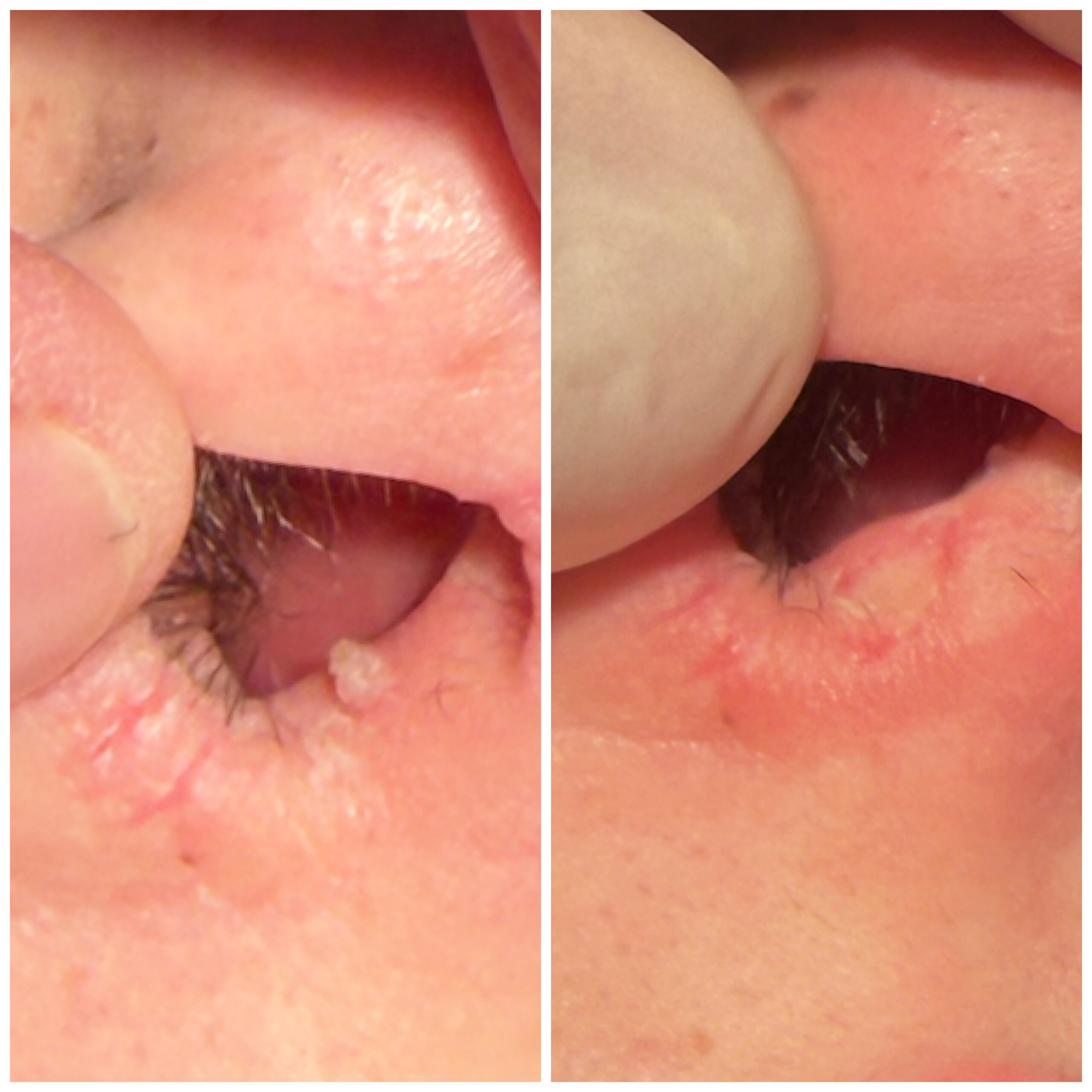
Few skin conditions cause more frustration than xanthelasma.
Although completely benign, these soft yellow plaques often appear on the upper or lower eyelids where they are highly visible. Many patients tell me they feel permanently tired or older than they really are because the lesions become the first thing people notice.
Fortunately, modern treatment can often achieve excellent cosmetic improvement.
What Is Xanthelasma?
Xanthelasma (pronounced zan-the-LAZ-ma) is the most common form of xanthoma, a harmless collection of cholesterol-rich cells that develops within the skin.
These deposits usually appear as soft, yellow or cream-coloured plaques around the eyelids, particularly close to the inner corners of the eyes.
They are completely benign.
Importantly, they are not skin cancer, they are not infectious and they cannot spread from one person to another.
Why Does Xanthelasma Develop?
The exact reason why xanthelasma develops is not always clear.
In some people, it is associated with raised cholesterol or other abnormalities of fat metabolism.
However, many patients with xanthelasma have completely normal cholesterol levels.
Other contributing factors may include:
- Increasing age.
- A family tendency.
- Diabetes.
- Thyroid disorders.
- Liver disease (less commonly).
For this reason, particularly when xanthelasma develops at a younger age or is extensive, I often recommend discussing a cholesterol profile and general cardiovascular risk assessment with your GP.
Are They Dangerous?
No.
Xanthelasma itself is entirely harmless.
The lesions do not become cancerous and usually cause no pain or discomfort.
For most people, the concern is cosmetic rather than medical.
However, because xanthelasma can occasionally act as a marker of underlying lipid abnormalities, appropriate blood tests may be worthwhile if these have not already been performed.
Can Creams Remove Xanthelasma?
Unfortunately, no.
Despite numerous products advertised online, there is currently no scientifically proven cream capable of removing established xanthelasma.
The cholesterol deposits lie beneath the surface of the skin and therefore cannot simply be dissolved by topical treatments.
Patients should be cautious of products making unrealistic claims.
How Is Xanthelasma Treated?
Several treatment methods are available, including:
- Surgical excision.
- Carbon dioxide (CO₂) laser.
- Chemical cautery using trichloroacetic acid (TCA) in selected cases.
- Radiofrequency surgery.
Each technique has advantages and limitations.
The most appropriate treatment depends upon the size, thickness and exact location of the lesions.
Why I Often Recommend Radiofrequency Surgery
In my practice, high-frequency radiofrequency (RF) surgery has become one of my preferred treatment options for carefully selected cases.
Using a very fine electrode, the abnormal tissue can be precisely ablated while minimising thermal damage to the surrounding healthy skin.
The advantages include:
- Excellent precision.
- Minimal bleeding.
- Good cosmetic healing.
- Rapid treatment.
- Usually performed under local anaesthetic.
- Often little downtime compared with more invasive procedures.
As with any procedure, no treatment is completely scar-free and careful aftercare is important to achieve the best possible cosmetic result.
Can Xanthelasma Return?
Unfortunately, yes.
Even after successful treatment, new cholesterol deposits may gradually develop over time.
Recurrence does not necessarily mean that treatment has failed.
Rather, it reflects an individual’s underlying tendency to form these deposits.
Maintaining a healthy lifestyle and addressing any underlying cholesterol abnormalities may reduce the likelihood of recurrence, although this cannot always prevent it completely.
What Happens During Treatment?
Following careful examination, the area is usually numbed with a small amount of local anaesthetic.
The xanthelasma is then precisely removed using radiofrequency surgery.
Most patients describe the procedure as surprisingly comfortable.
After treatment, a small superficial wound forms which gradually heals over the following one to two weeks.
Mild redness may persist for several weeks before blending into the surrounding skin.
Careful aftercare and protection from excessive sun exposure help optimise the cosmetic result.
Dr Kitchen’s Clinical Pearl
One of the most rewarding aspects of treating xanthelasma is seeing how such a relatively small procedure can restore confidence.
Patients often tell me that they had become increasingly aware of people looking at the yellow patches rather than making eye contact.
Although xanthelasma is medically harmless, its psychological impact should never be underestimated.
Successful treatment is not simply about removing a lesion.
It is about helping people feel comfortable in their own skin again.
Should I Have My Cholesterol Checked?
- First episode of xanthelasma under the age of 50 ✔️
- Strong family history of heart disease ✔️
- Diabetes ✔️
- Thyroid disease ✔️
- Previous high cholesterol ✔️
Although many people with xanthelasma have normal cholesterol levels, a simple blood test can be worth while and may identify treatable cardiovascular risk factors.
17. Xanthelasma/ Xanthelasmata almost ALWAYS appear around the eyelids
Xanthelasma Removal – My Experiences
I wanted to share my Xanthelasma experience with you which hopefully you will find useful. I really wish someone gave me this advice when I was researching Xanthelasma as it would have saved me a lot of time. However I guess all the research has sort of made me an “expert” so I feel good having done all the work nonetheless.
I had these ugly bumps called Xanthelasma around my eyes for years. I thought about removing them several years ago but a couple things caused me to rethink removal. First, it wasn’t covered by insurance which bothered me. They claim its “cosmetic”. It’s not like I’m getting a facelift. There are actual ugly bumps that seem to continue to grow around my eyes! How is that “cosmetic”? They are abnormal growths! Regardless, the main reason I postponed doing anything about removing them was because the option I was contemplating was having them surgically removed. I felt that a surgeon taking a knife and cutting out these little things was sort of overkill and dangerous. They cut big incisions around the eyes, they would have to stitch them up afterwards and then you have to hope they heal without scars. No way was I going to do that. I felt that there had to be a better, safer and frankly less uncomfortable way.
So I researched on and off. I would spend more time researching after I noticed someone looking at them closely while I talk to them. Really uncomfortable. So finally, I decided I was going to dive into the research and just get them removed once and for all.
I researched the many different ways to remove Xanthelasma and one particular procedure caught my attention. It was called “Radiosurgery”. Radiosurgery does not involve a knife cutting into your skin. Instead it’s a non-pressure, smooth, incision that produces no blood. Sort of like a laser but with less heat and tissue alteration (I’ve had to do some cutting and pasting from some materials I read to give you this description). The doctor can easily hold the Radiosurgery device…sort of like a small pencil with a small laser as a tip. It cuts the skin without a blade thus less risky. There are no sutures or stitches afterward either. There is some swelling but some ice packs after surgery help keep that in line.
I was living on the west coast at the time so I researched dermatologists that have Radiosurgery machines in the offices. I found one and set up a consultation. They said they can do the procedure. I was a little nervous because this particular doctor never used the Radiosurgery device on Xanthelasma before. But she seemed competent and careful so I agreed to do it.
I came in the office on the day of surgery, they laid me down (face up) on a table, and they injected my eyelids with something that numbs them so I don’t feel anything around my eyes. Then she took this little pencil and slowly and gently cut these things out of my eyelids. I felt no pain except I did feel the heat a little but wasn’t painful. I was wide awake during everything but I didn’t feel any pain at all. 1It was definitely awkward and uncomfortable having this procedure happening so darn close to my eyes. But I was glad she wasn’t using a knife!
After about 45 minutes or so the first procedure was done. I had the procedures done over 2 visits as I had Xanthelasma on both eyes, both above and below. So we did the removal over two visits. I could easily have had them all removed at once but I liked being able to see how the healing process of the first part goes before doubling down.
Immediately after the surgery my eyes definitely looked pretty bad. But remarkably, the healing process was pretty quick. I had the procedure done on a Saturday, and by Monday the healing process kicked in so much that when I went to the office, no one noticed the redness. Granted I didn’t have a lot of close conversations at the office on Monday, but I had meetings and no one said things like “who punched you in the face”? So those made me feel better. I’m guessing that if someone had very large Xanthelasmata then maybe it would take longer to heal and more noticeable post operation. Mine weren’t too big so I guess that helped. After a week or so any swelling, scabbing etc was gone. The only thing that took awhile to adjust was the skin colouration in that area. I have a little darker pigment (my Italian heritage) and the area where the Xanthelasma was removed healed with the skin turning lighter. Over time the colour blended in and looked more normal, but when I look closely I can tell that the skin is a slightly different shade, but totally unnoticeable to anyone except me who has been looking at the same face in the mirror for the last 40 years.
Oh yeah, the second procedure came out fine too, and they even corrected something that was missed in the first procedure (she didn’t go deep enough on one of the xanthelasmata).
Ok, I know this is a long post but I just wanted to end this my saying that I’M VERY SATISFIED WITH THE REMOVAL OF THE XANTHELASMA AND SO MUCH HAPPIER THAT I DON’T HAVE TO SEE THOSE UGLY BUMPS ANYMORE.
I feel strongly that you should not get traditional surgery so stay away from the surgeon. Everyone can have their own opinion, but based on the years of research that I’ve done and numerous consultations, I have a very strong viewpoint that for the removal of Xanthelasma, Radiosurgery is the best method for removal for just about every reason (it works, easy, not that expensive compared to surgery, no stitches, safer, no knives, good healing process etc).
I had the procedure done over a year ago and I just wanted to share my experiences as I wish someone gave me this advice when I was looking for removal options.
Good luck and I hope this post helps you.
AC_Warwickshire
18. Precision RadioFrequency Surgery for Benign Skin Lesions
What is radiofrequency surgery?
It uses very high-frequency alternating electrical energy delivered through a fine electrode to cut or remove tissue with exceptional precision.
Why Is Radiofrequency Surgery Different?
There are several well-established methods of removing benign skin lesions, and each has its place in modern medical practice. The most appropriate treatment depends upon the type of lesion, its size, its location and the cosmetic outcome that both doctor and patient hope to achieve.
One of the questions I am frequently asked is, “Why do you use radiofrequency surgery instead of simply cutting it off?”
The answer lies in the precision of the technology.
Scalpel Surgery
Traditional surgical excision using a scalpel remains an excellent treatment for many skin lesions and is often the preferred option when a lesion requires complete removal for laboratory examination (histology) or when deeper tissue needs to be excised.
However, scalpel surgery usually involves a larger incision and often requires stitches. Healing may therefore take a little longer, and a linear scar is usually inevitable.
Cryotherapy (Freezing)
Cryotherapy removes tissue by freezing it with liquid nitrogen.
It is quick and convenient for selected lesions, particularly certain viral warts and some superficial sun-damaged skin.
However, the depth of freezing can be less predictable than other techniques, and temporary blistering, pigment changes or the need for repeat treatments may occur.
Laser Surgery
Lasers use concentrated light energy to remove or vaporise tissue.
They are excellent for certain skin conditions and can produce outstanding cosmetic results when used appropriately.
However, laser equipment is expensive, and not every lesion is best treated with a laser. In many cases, radiofrequency surgery offers similar precision with less collateral thermal damage and greater versatility.
Electrocautery
Although often confused with radiofrequency surgery, electrocautery works in a different way.
Electrocautery uses a heated metal tip to burn tissue.
Radiofrequency surgery, by contrast, generates heat within the tissue itself using very high-frequency alternating electrical energy delivered through a fine electrode.
Because the electrode itself remains relatively cool, there is generally less unnecessary thermal damage to surrounding tissue, allowing more precise treatment and often better cosmetic healing.
Why I Often Choose Radiofrequency Surgery
For many benign skin lesions, radiofrequency surgery combines exceptional precision with excellent cosmetic outcomes.
It allows tissue to be removed accurately while minimising trauma to the surrounding healthy skin.
For this reason it has become one of my preferred techniques for treating many facial lesions where cosmetic appearance is particularly important.
⸻
What Happens During Treatment?
One of the commonest concerns patients have is not about the treatment itself, but about what to expect on the day.
Fortunately, radiofrequency surgery is usually a straightforward outpatient procedure.
Following a careful examination and confirmation that radiofrequency surgery is the most appropriate treatment, the area is cleaned thoroughly and a small amount of local anaesthetic is administered. Most patients describe this as the only mildly uncomfortable part of the procedure, lasting just a few seconds.
Once the area is numb, treatment can begin immediately.
Using a very fine radiofrequency electrode, the lesion is carefully removed with great precision. Because radiofrequency energy seals tiny blood vessels as it works, bleeding is usually minimal, providing a clear view throughout the procedure.
Many lesions can be removed without the need for stitches, particularly smaller superficial growths such as skin tags, seborrhoeic keratoses and selected facial lesions.
After treatment, the area is gently cleaned and, where appropriate, covered with a small dressing. Detailed aftercare advice is provided before you leave the clinic.
Most patients are able to return home immediately and resume normal daily activities, although specific advice will depend upon the type and location of the lesion treated.
⸻
What Conditions Can Be Treated?
Radiofrequency surgery is suitable for a wide range of benign skin lesions, including carefully selected:
- Skin tags
- Seborrhoeic keratoses
- Warts
- Verrucae
- Xanthelasma
- Benign moles (where clinically appropriate)
- Certain cysts
- Small benign facial lesions
- Cherry angiomas
- Fibrous papules
- Selected benign vascular lesions
Not every skin lesion is suitable for radiofrequency treatment, which is why a careful clinical assessment is always the first step.
⸻
Does It Hurt?
Most patients are pleasantly surprised by how comfortable the procedure is.
The local anaesthetic used to numb the area may cause a brief stinging sensation lasting only a few seconds.
Once the anaesthetic has taken effect, treatment itself is usually painless.
Following the procedure, some patients experience mild tenderness similar to a grazed knee or superficial cut for a few days. This is usually well controlled with simple pain relief if required.
⸻
Will I Have a Scar?
Whenever the skin is treated, some degree of healing response will occur.
The aim of modern skin surgery is therefore not to promise no scar, but to achieve the best possible cosmetic scar.
Radiofrequency surgery allows tissue to be removed with exceptional precision while minimising unnecessary trauma to surrounding skin.
For many benign lesions—particularly on the face—this often results in excellent cosmetic healing.
Scar appearance depends upon several factors, including:
- The size and depth of the lesion.
- Its location on the body.
- Individual healing characteristics.
- Sun exposure during healing.
- Following the recommended aftercare instructions.
Most scars continue to mature and improve over several months.
⸻
Advantages of Radiofrequency Surgery
Why many patients choose radiofrequency treatment
✓ Exceptional precision.
✓ Minimal bleeding.
✓ Excellent cosmetic healing.
✓ Usually performed under local anaesthetic.
✓ Often no stitches required.
✓ Rapid outpatient procedure.
✓ Suitable for many delicate facial areas.
✓ Minimal disruption to surrounding healthy tissue.
✓ Tissue can often be sent for histological examination where clinically appropriate.
✓ Patients can usually return home immediately after treatment.
⸻
Why Dr Kitchen Prefers Radiofrequency Surgery
Throughout my career I have always believed that selecting the right treatment is just as important as performing it well.
Over many years I have used a variety of techniques, including conventional surgery, cryotherapy and laser treatments. Each has an important role in modern skin surgery.
For many benign skin lesions, however, I have found high-frequency radiofrequency surgery to offer an excellent balance between precision, safety and cosmetic outcome.
For many years I have used the Ellman Surgitron system, a respected medical device that delivers high-frequency radiofrequency energy with remarkable accuracy. Its precision allows me to treat many lesions while preserving as much healthy surrounding tissue as possible—something that is particularly important when working on the face, eyelids and other cosmetically sensitive areas.
Technology alone, however, does not determine the outcome.
Successful treatment depends upon accurate diagnosis, careful patient selection, meticulous technique and clear communication about what can realistically be achieved.
Dr Kitchen’s Clinical Pearl
“After nearly four decades in practice, I’ve learnt that patients rarely ask me, ‘What’s the newest machine?’ They ask, ‘What would you recommend if I were your family?’ My answer is always based on experience rather than fashion. Radiofrequency surgery has earned its place in my practice because, in carefully selected cases, it consistently delivers the combination of precision, safety and cosmetic results that both my patients and I value.”
More about Mole Mapping
Testimonials
“The procedure itself is relatively pain-free and very quick
I had mole removal treatments 4 months ago and wanted to say the results are really good. The procedure itself is relatively pain-free and very quick. No stitches which are great. Extremely professional and friendly atmosphere in the clinic. Highly recommended to anyone considering. Thanks again to Hugo and his team!
Edward”
“Wow what a clinic! I took my daughter who was so scared about having 2 warts removed on her toe and the doctor was superb making my daughter laugh and totally taking her mind off it. We had this clinic recommended to us and I can see why now. Superb service, a massive thank you.
SC”
“Dear Dr Hugo Kitchen,
Thank you so much for removing the mole on my neck before I went to uni, I’m so happy with it. It has healed really well and there is only a faint mark mark which is remarkable considering how recently the removal happened.
I cannot thank you enough for carrying out the procedure, I cannot explain to you how pleased I am with it.
Thank you again,
L W”
“Hi Hugo,
Thank you so much for removing my moles.
I am thrilled to bits with the results..Absolutely amazing!
3 weeks on and they have virtually disappeared.
Kind regards
Jacky x”
“To the Stratford Dermatherapy Clinic
I had moles!!!….no, not in my garden, on my face. Having finally plucked up courage I made an appointment visit the Stratford Dermatherapy Clinic for a consultation. The initial consultation with Dr Hugo was very thorough, giving me lots of information and an opportunity to ask any questions. Dr Hugo talked though all my options and explained the chosen procedure. The consultation was free and there was no pressure to continue if I had not wished to. I chose to have my moles removed by a high tech gadget that removed them with minimal fuss, no pain and fabulous results. I am very pleased that I went ahead with the procedure and would recommend Dr Hugo for anyone who has thought about facial mole removal.
Thank you so much Dr Hugo and your clinic
Kind regards
Amanda Austen-Jones”
Mole removal Warwickshire at Stratford Dermatherapy Clinic
- Moles, skin tags and polyps, warts, and cysts can be removed for cosmetic reasons and to improve aesthetic appearance.
- Quicker recovery time.
- Consultation, diagnosis and removal of moles are carried in one appointment.
- Fast diagnosis carried out by histologists for moles.
- Appointments available six days a week including evenings.
- Same or next day appointments for Mole Removal @ Stratford Dermatherapy Clinic, Warwickshire can be booked.
See more before and afters in our gallery
What Happens During Your Mole Assessment?
For many people, arranging a mole assessment can feel daunting. It is perfectly natural to wonder what the examination involves, whether it will be uncomfortable, or whether you might receive unexpected news.
My aim is always to make the consultation as relaxed, informative and reassuring as possible.
Step 1 – Listening to Your Concerns
Every consultation begins with a conversation.
I want to understand why you have come to see me.
Perhaps you have noticed a mole changing in colour or size. Maybe a partner has pointed out a lesion on your back, or your hairdresser has noticed something on your scalp. You may simply want reassurance because skin cancer runs in your family.
There are no unimportant questions.
Understanding your concerns is often just as important as examining the lesion itself.
Step 2 – Taking a Brief Medical History
Before examining your skin, I will ask a few simple questions, including:
- How long have you noticed the lesion?
- Has it changed in size, colour or shape?
- Has it become itchy, painful or started bleeding?
- Have you ever had skin cancer before?
- Is there a family history of melanoma?
- Have you experienced significant sun exposure or severe sunburn, particularly during childhood?
These questions help me assess your individual level of risk and place any findings into the correct clinical context.
Step 3 – Careful Clinical Examination
The mole or skin lesion is then examined under good lighting.
At this stage, I assess its size, shape, colour, border and overall appearance. I also compare it with the surrounding skin and, where appropriate, with your other moles.
Sometimes the answer is immediately obvious.
Occasionally, a closer examination is required.
Step 4 – Dermoscopic Assessment
One of the most valuable tools in modern dermatology is the dermatoscope.
This specialised instrument allows me to examine structures beneath the surface of the skin that cannot be seen with the naked eye.
The examination is completely painless and takes only a few moments, but it provides an extraordinary amount of additional information that helps distinguish many benign lesions from those requiring further investigation.
Step 5 – Digital Imaging and AI Analysis (Where Appropriate)
For suitable lesions, high-quality digital images may be taken and analysed using advanced artificial intelligence software.
The AI compares the appearance of the lesion with a vast database of previously assessed skin lesions, providing an additional level of analysis to support clinical decision-making.
It is important to remember that this technology is an aid to diagnosis rather than a replacement for clinical judgement. Every result is interpreted alongside your medical history, examination findings and my own experience.
Step 6 – Explaining the Findings
One of the most important parts of any consultation is explaining exactly what I have found.
I believe patients should leave understanding their diagnosis rather than simply being told a result.
Where possible, I will show you the dermoscopic images, explain the features I can see and discuss why I believe a lesion is benign or why I recommend further investigation.
Most patients tell me this is the most reassuring part of the consultation.
Knowledge removes uncertainty.
Step 7 – Agreeing the Best Management Plan
If the lesion is entirely benign, you may simply require reassurance and advice about self-monitoring.
If removal is appropriate, we will discuss the available treatment options, including the likely cosmetic outcome, healing process and aftercare.
If I believe a lesion requires urgent investigation within the NHS or by a specialist skin cancer service, I will explain exactly why and ensure you understand the next steps.
Whatever the outcome, my aim is that you leave the consultation with clarity, confidence and a clear plan.
Dr Kitchen’s Clinical Pearl
Over nearly four decades, I have learnt that patients are rarely frightened by the examination itself. What causes the greatest anxiety is not knowing what to expect.
By taking the time to explain each step, answer every question and discuss the findings openly, most people leave feeling reassured—even when further treatment is required.
Good medicine is not simply about making the correct diagnosis.
It is about helping people understand it.
What happens during the treatment?
The area to be treated will be cleaned and might be numbed with local anaesthetic. The removal will then take place and be carried out in one of two ways:
Micro-excision: the mole will be shaved off either at or slightly below the level of skin. No stitches are used so any bleeding will be stopped by either cauterising the area.
Excision using the ELLMAN SURGITRON Radio-Frequency device is the treatment method of choice as it is clean and immediate with no bleeding.
What happens after the treatment?
There isn’t usually a requirement for a follow-up appointment when there has been a simple mole procedure. However we do like to check up at 4 weeks to ensure the removal site is healing well. Some moles removed are sent for a lab analysis and if there are any unusual features you will be contacted by telephone.
If there are stitches, eg after excision of a large cyst, these will be removed within 5-7 days if on the face and within 8-21 days for stitches elsewhere.
To prevent infection the area should be kept clean and no unnecessary or stress should be placed on the wound, to allow it adequate time to heal.
19. Skin Cancer and Pre-Cancerous Skin Lesions:-
A complete patient guide:-
Understanding Moles – The Science Without the Scare
Before we discuss the warning signs of melanoma, it’s worth understanding what a mole actually is. I often find that once patients understand the biology, much of their anxiety disappears.
A mole, or melanocytic naevus to give it its medical name, is simply a cluster of pigment-producing cells called melanocytes. These cells are responsible for making melanin, the pigment that gives our skin, hair and eyes their colour and provides some protection from ultraviolet radiation.
For reasons that are not yet completely understood, these cells sometimes gather together into small nests rather than remaining evenly distributed throughout the skin. The result is what we recognise as a mole.
Some babies are born with moles, although most appear during childhood and adolescence. It is perfectly normal for new moles to develop into your twenties and, occasionally, your thirties. Hormonal changes during puberty or pregnancy may also cause existing moles to darken slightly or become more noticeable. These changes are often entirely physiological.
Indeed, if you looked closely at the skin of ten healthy adults, you would probably find that every one of them has a unique pattern of moles. Some people have only a handful, while others may have well over one hundred. Genetics plays a major role, but lifetime sun exposure also contributes.
This is one of the reasons why there is no such thing as a “normal” mole. Moles come in an astonishing variety of colours, shapes and sizes. Some are completely flat, others are raised. Some are flesh coloured, others are light brown, dark brown or almost black. Many have tiny hairs growing through them. Contrary to popular belief, hairs growing from a mole are usually a reassuring rather than an alarming feature.
Patients often ask me, “Doctor, why has this mole suddenly appeared?”
In many cases, there is no sinister explanation whatsoever. The skin is a remarkably dynamic organ. New moles can develop naturally, particularly in younger adults. Seborrhoeic keratoses—one of the commonest benign skin growths—are frequently mistaken for new moles. Sun damage accumulated over decades can also produce lesions that patients suddenly notice one morning despite them having evolved slowly over several years.
The important point is this: finding a new mole does not automatically mean skin cancer.
Equally, having a mole for twenty or even thirty years does not guarantee that it should never be examined if it begins to change. Medicine is rarely black and white. It is about recognising patterns, identifying exceptions and knowing when further assessment is sensible.
One of the greatest misconceptions I encounter is the belief that melanoma always develops from an existing mole. In reality, many melanomas arise as entirely new pigmented lesions rather than evolving from a mole that has been present for years. That is why becoming familiar with your skin as a whole is often more valuable than obsessively monitoring one individual mole.
Another myth is that every dark mole is dangerous. Again, this is simply untrue. Some of the darkest moles I see prove to be completely benign, while occasionally a melanoma may be surprisingly pale. Colour alone tells only part of the story.
So what am I looking for when I assess a patient?
Firstly, I take a careful history. Has the lesion changed? Over what period? Is it itching, bleeding, crusting or failing to heal? Is there a personal or family history of melanoma? Has there been significant childhood sunburn? Has the patient noticed similar lesions elsewhere?
Only then do I begin the physical examination.
To the naked eye I assess symmetry, borders, colour and overall appearance. I then examine the lesion using dermoscopy—a specialised magnifying instrument with polarised light that allows me to visualise pigment networks, blood vessels and structures hidden beneath the skin surface. It is one of the most valuable advances in skin cancer diagnosis over the past few decades.
More recently, artificial intelligence has become another useful tool in clinical practice. Used responsibly, AI can compare dermoscopic images with thousands of documented lesions, offering an additional opinion that complements clinical judgement. However, I regard AI as exactly that—an assistant. It can never replace a thorough history, careful examination and the experience gained from seeing many thousands of lesions over nearly four decades.
Patients are often surprised when I tell them that I spend as much time reassuring people as I do referring them. Thankfully, the overwhelming majority of lesions I assess are benign. Sometimes all that is required is explanation, education and the confidence that comes from knowing an experienced clinician has examined the lesion carefully.
That reassurance has genuine value. Anxiety itself can become a burden, and one of my aims has always been to replace uncertainty with understanding.
Of course, there are occasions when I recommend removal or referral. If a lesion demonstrates suspicious dermoscopic features, is changing significantly or simply cannot be confidently classified as benign, then further investigation is the correct course of action. Modern medicine is at its best when it combines vigilance with common sense—not unnecessary alarm, but not false reassurance either.
Over the years I have often reflected that patients rarely remember the technical details of a consultation. What they remember is how they felt when they left the room. Did they feel listened to? Did they understand what had been explained? Were they reassured without being dismissed?
That has always been my philosophy.
Knowledge should replace fear, but knowledge delivered with kindness is even more powerful.
20. Basal Cell Carcinoma (BCC) The Commonest Skin Cancer:-
Few words cause more anxiety than “skin cancer.” Fortunately, not all skin cancers behave in the same way, and one of the commonest forms—Basal Cell Carcinoma (BCC)—is also one of the most treatable when recognised early.
A Basal Cell Carcinoma is the most frequently diagnosed skin cancer in the United Kingdom. It develops from the basal cells found in the deepest layer of the epidermis, the outer layer of the skin.
Unlike melanoma, BCC almost never spreads (metastasises) to distant parts of the body. Instead, it grows slowly and tends to invade the surrounding skin and underlying tissues if left untreated. This is why early diagnosis and treatment are important. Although it is rarely life-threatening, delaying treatment may result in a larger operation and a more noticeable scar.
What Causes Basal Cell Carcinoma?
The single greatest risk factor is long-term exposure to ultraviolet (UV) radiation, whether from natural sunlight or artificial sources such as sunbeds.
BCCs are therefore more common on areas that receive the greatest lifetime sun exposure, including:
- The nose
- Eyelids
- Forehead
- Ears
- Cheeks
- Neck
- Scalp (particularly in balding men)
Fair-skinned individuals, those who burn easily, outdoor workers and people with a history of frequent sun exposure are at increased risk. However, BCC can develop in anyone.
What Does a Basal Cell Carcinoma Look Like?
One reason BCC is sometimes missed is that it does not always resemble the dramatic images people associate with skin cancer.
It may appear as:
- A small pearly or shiny bump.
- A pink patch that slowly enlarges.
- A sore that repeatedly heals before breaking down again.
- A lesion with tiny visible blood vessels (telangiectasia).
- A persistent ulcer that fails to heal.
- A waxy scar-like area, particularly in less common forms.
Many patients tell me, “I thought it was just a spot that wouldn’t go away.” That is a remarkably common story.
How Is It Diagnosed?
Diagnosis begins with a careful clinical examination.
At Stratford Dermatherapy Clinic I routinely examine suspicious lesions using dermoscopy, which allows structures beneath the surface of the skin to be visualised far more clearly than with the naked eye.
Where appropriate, digital photography and AI-assisted assessment may provide additional information, although technology never replaces clinical judgement.
If there is any concern that a lesion represents a skin cancer, referral for biopsy or definitive treatment is arranged in accordance with current clinical guidance.
How Is It Treated?
Treatment depends upon the size, location and subtype of the tumour.
Options may include:
- Standard surgical excision.
- Mohs micrographic surgery for selected facial lesions.
- Curettage and cautery for suitable superficial lesions.
- Topical treatments for carefully selected superficial BCCs.
- Radiotherapy in selected circumstances.
The most appropriate treatment is determined after careful assessment.
Can Basal Cell Carcinoma Be Prevented?
Not every BCC can be prevented, but the risk can be reduced by sensible sun protection throughout life.
This includes:
- Avoiding prolonged exposure during the strongest midday sun.
- Wearing protective clothing and wide-brimmed hats.
- Using a broad-spectrum sunscreen appropriate for your skin type.
- Avoiding sunbeds.
- Becoming familiar with your own skin and seeking advice if a lesion changes or fails to heal.
Dr Kitchen’s Clinical Pearl
One of the commonest comments I hear is:
“I didn’t think it could be skin cancer because it never hurt.”
Basal Cell Carcinomas are often painless.
In fact, one of their defining features is that they can quietly enlarge over many months while causing little or no discomfort.
Any sore, lump or patch of skin that persists for several weeks without healing deserves assessment—not because it is likely to be serious, but because it is always better to know than to worry.
21. Squamous Cell Carcinoma (SCC)_Why Early treatment matters.
Whilst Squamous Cell Carcinoma (SCC) is less common than Basal Cell Carcinoma, it remains one of the most frequently diagnosed skin cancers in the UK and deserves prompt medical attention.
Unlike Basal Cell Carcinoma, which very rarely spreads elsewhere in the body, Squamous Cell Carcinoma has a greater potential to invade deeper tissues and, in a small proportion of cases, spread to nearby lymph nodes or other parts of the body if left untreated.
The reassuring news is that, when diagnosed early, the vast majority of Squamous Cell Carcinomas are treated very successfully.
The key is recognising the warning signs and seeking assessment without unnecessary delay.
What Causes Squamous Cell Carcinoma?
The most important cause is long-term cumulative exposure to ultraviolet (UV) radiation.
Years of sun exposure gradually damage the DNA within skin cells. Over time, these changes can cause the squamous cells within the epidermis to grow in an uncontrolled way.
Other recognised risk factors include:
- Fair skin that burns easily.
- Increasing age.
- A history of outdoor occupations or hobbies.
- Previous skin cancers.
- Weakened immune systems, including some transplant recipients.
- Long-term sunbed use.
- Chronic wounds or scars (less commonly).
Most SCCs develop on areas that have received the greatest lifetime sun exposure, particularly the:
- Face
- Ears
- Lips
- Scalp
- Neck
- Forearms
- Back of the hands
What Does Squamous Cell Carcinoma Look Like?
Unlike melanoma, Squamous Cell Carcinoma often develops gradually.
It may appear as:
- A firm, rough or scaly lump.
- A persistent crusted patch.
- A sore that fails to heal.
- A lesion that repeatedly bleeds after minor trauma.
- A rapidly enlarging tender nodule.
- A thickened, wart-like growth.
Many patients describe the lesion as “a sore that just won’t heal.”
That simple description should never be ignored.
Does Squamous Cell Carcinoma Hurt?
Not always.
Some SCCs are completely painless during their early stages.
Others may become tender, sore or bleed easily, particularly as they enlarge.
The absence of pain should never be taken as reassurance that a lesion is harmless.
How Is It Diagnosed?
Careful clinical examination is always the first step.
At Stratford Dermatherapy Clinic I assess suspicious lesions using dermoscopy, which allows subtle surface and vascular features to be examined in far greater detail than is possible with the naked eye.
Where appropriate, digital photography and AI-assisted analysis may provide additional information. However, no technology replaces careful clinical judgement and experience.
If there is any suspicion that a lesion may represent Squamous Cell Carcinoma, I arrange prompt referral for biopsy and definitive treatment in accordance with current clinical guidance.
How Is Squamous Cell Carcinoma Treated?
Treatment depends upon the size, depth and location of the tumour.
Options commonly include:
- Surgical excision.
- Mohs micrographic surgery for selected cases.
- Radiotherapy where appropriate.
- Specialist management for more advanced disease.
Early treatment offers the highest chance of complete cure while minimising the amount of surgery required.
Can Squamous Cell Carcinoma Be Prevented?
Although not every case can be prevented, sensible sun protection significantly reduces future risk.
Practical measures include:
- Avoiding excessive ultraviolet exposure.
- Wearing protective clothing and hats.
- Applying broad-spectrum sunscreen when appropriate.
- Avoiding sunbeds.
- Regularly examining your own skin.
- Seeking medical advice if a persistent lesion develops.
Individuals who have already had one skin cancer are at increased risk of developing further lesions and should remain particularly vigilant.
When Should I Seek Medical Advice?
Arrange an assessment if you notice any skin lesion that:
- Persists for more than four to six weeks.
- Continues to bleed or crust.
- Is enlarging.
- Feels unusually firm.
- Develops on chronically sun-damaged skin.
- Simply concerns you.
The vast majority of suspicious lesions prove to be treatable, and many turn out not to be cancer at all. However, it is always better to investigate a persistent lesion than to ignore it.
Dr Kitchen’s Clinical Pearl
One lesson that nearly four decades in practice has taught me is that patients often wait because they hope a lesion will simply disappear.
Occasionally it does.
But when a sore, crust or lump remains unchanged for several weeks—or slowly becomes larger—it is telling us something.
Most people are relieved to discover that the diagnosis is less serious than they feared.
For the smaller number in whom a Squamous Cell Carcinoma is diagnosed, that decision to seek advice early often makes treatment considerably simpler and leads to an excellent outcome.
22. Actinic Keratoses_Sun damage that shouldn’t be ignored.
If you have ever noticed a rough, dry patch of skin that simply refuses to disappear, you may be looking at an Actinic Keratosis (AK).
These small, sun-damaged areas of skin are extremely common, particularly as we get older. Although many remain stable for years, they are considered pre-cancerous lesions, meaning they have the potential to develop into Squamous Cell Carcinoma (SCC) if left untreated.
Fortunately, most Actinic Keratoses can be recognised early and treated effectively, helping to reduce that risk.
What Causes Actinic Keratoses?
Actinic Keratoses develop after years of cumulative exposure to ultraviolet (UV) radiation from the sun.
Unlike sunburn, which occurs after excessive exposure over a short period, AKs reflect the gradual effects of sunlight accumulated over many years.
This explains why they are particularly common in:
- Fair-skinned individuals.
- People over the age of 50.
- Anyone who has enjoyed a lifetime of outdoor activities.
The face, scalp, ears, forearms, backs of the hands and lower legs are the areas most commonly affected because they receive the greatest lifetime sun exposure.
What Do They Look Like?
Many patients are surprised that Actinic Keratoses are often felt before they are seen.
The earliest sign may simply be a small rough patch that feels like fine sandpaper when you run your finger across the skin.
As they develop, they may become:
- Pink, red or skin-coloured.
- Persistent despite moisturisers.
Some remain tiny for years, while others slowly enlarge.
Unlike eczema or dry skin, they rarely disappear completely without treatment.
Are They Dangerous?
Most Actinic Keratoses never become cancerous.
However, because they represent abnormal sun-damaged skin cells, they are regarded as pre-cancerous lesions.
A small proportion will progress to Squamous Cell Carcinoma over time, although it is impossible to predict which individual lesion may do so.
For this reason, persistent or changing AKs should be assessed by an experienced clinician.
Early recognition and treatment are usually straightforward and may help prevent future complications.
How Are Actinic Keratoses Diagnosed?
Diagnosis is usually made following careful clinical examination.
At Stratford Dermatherapy Clinic I routinely use dermoscopy to examine suspicious lesions in greater detail, allowing me to distinguish many Actinic Keratoses from other common skin conditions.
Occasionally, if the diagnosis is uncertain or a lesion shows features suggestive of early Squamous Cell Carcinoma, a biopsy or referral for further assessment may be recommended.
How Are They Treated?
Treatment depends upon:
- Individual patient factors.
Common treatment options include:
- Prescription topical creams or gels.
- Photodynamic Therapy (PDT).
- Curettage in selected cases.
- Surgical removal where appropriate.
The most suitable treatment is chosen after careful assessment of the individual lesion and the surrounding skin.
In addition to treating visible lesions, ongoing sun protection plays an important role in reducing the development of further Actinic Keratoses.
Can They Be Prevented?
While existing sun damage cannot be reversed completely, future damage can often be reduced.
Simple measures include:
- Avoiding excessive midday sun.
- Wearing wide-brimmed hats and protective clothing.
- Applying a broad-spectrum sunscreen appropriate for your skin type.
- Becoming familiar with your own skin and arranging regular skin examinations if you have extensive sun damage.
When Should I Seek Medical Advice?
Arrange an assessment if you notice:
- A rough patch that persists for several weeks.
- A lesion that becomes thicker or rapidly enlarges.
- Persistent crusting or bleeding.
- A lesion that simply concerns you.
Although many rough patches prove to be harmless, persistent sun-damaged lesions deserve professional assessment.
Dr Kitchen’s Clinical Pearl
One of the commonest comments I hear is:
“I thought it was just dry skin.”
Sometimes it is.
But persistent rough patches that repeatedly return despite moisturisers deserve a closer look.
Most Actinic Keratoses are easily treated, and many patients are relieved to discover that treatment at this stage is often much simpler than waiting until more significant changes have developed.
Protecting your skin from further sun damage is one of the best investments you can make—not only for cosmetic reasons, but for your long-term skin health.
23. Cherry Angiomas – Small Red Spots That Are Usually Harmless
Many people notice the gradual appearance of small, bright red spots on their skin as they get older. These tiny lesions are known as Cherry Angiomas (sometimes called Campbell de Morgan spots or senile angiomas) and are one of the most common benign skin growths seen in adults.
Although their appearance can be alarming, particularly if they develop suddenly or begin to bleed after being caught on clothing, Cherry Angiomas are almost always completely harmless and are not a form of skin cancer.
They are made up of tiny clusters of dilated blood vessels within the skin, giving them their characteristic bright red or purple colour.
What Causes Cherry Angiomas?
The exact cause is not fully understood, but Cherry Angiomas become increasingly common with age.
They are thought to develop as part of the natural ageing process of the skin and its small blood vessels.
Several factors may contribute, including:
- Increasing age.
- Genetic predisposition.
- Hormonal influences.
- Natural changes within the skin over time.
Most people begin to notice them from their thirties or forties onwards, and many individuals eventually develop dozens of them.
They are not contagious and are not caused by poor health or lifestyle choices.
What Do Cherry Angiomas Look Like?
Cherry Angiomas are usually easy to recognise.
They typically appear as:
- Small, round or oval red spots.
- Bright cherry-red, crimson or purple in colour.
- Smooth and slightly raised.
- Usually between 1 and 5 millimetres in diameter.
- Soft to the touch.
- Found most commonly on the trunk, chest, abdomen, back and arms.
Some remain tiny throughout life, while others gradually enlarge into small dome-shaped bumps.
Because they contain numerous small blood vessels, they may bleed surprisingly easily if accidentally scratched or caught.
Are Cherry Angiomas Dangerous?
No.
Cherry Angiomas are benign vascular lesions and do not become skin cancer.
However, if a new red lesion appears and behaves differently—for example, enlarging rapidly, changing colour, becoming persistently ulcerated or bleeding repeatedly without obvious injury—it should always be assessed to confirm the diagnosis.
Although Cherry Angiomas are usually straightforward to identify, not every red skin lesion is an angioma.
How Are Cherry Angiomas Diagnosed?
Diagnosis is usually made during a careful clinical examination.
At Stratford Dermatherapy Clinic I routinely use dermoscopy, allowing the tiny blood vessel patterns within the lesion to be examined in detail.
In the vast majority of cases, this confirms the diagnosis immediately and provides reassurance that no further investigation is required.
Occasionally, if a lesion has unusual features or the diagnosis is uncertain, further assessment or biopsy may be recommended.
Do They Need Treatment?
In most cases, no treatment is medically necessary.
Many patients simply choose to leave them alone.
Treatment may be considered if a Cherry Angioma:
- Frequently catches on clothing or jewellery.
- Bleeds repeatedly.
- Has become irritated.
- Is cosmetically bothersome.
- Causes anxiety because of its appearance.
How Are Cherry Angiomas Removed?
Several treatment methods are available, including:
- Radiofrequency surgery.
- Intense Pulsed Light (IPL)
- Electrocautery.
- Laser treatment.
- Cryotherapy in selected cases.
- Surgical excision for unusual lesions.
The most appropriate treatment depends upon the size, location and appearance of the lesion.
One of the advantages of modern radiofrequency surgery is that it allows very precise removal while minimising damage to the surrounding skin. Healing is generally rapid, and most patients are delighted with the cosmetic outcome.
Will They Come Back?
Once an individual Cherry Angioma has been completely removed, it usually does not return.
However, because they are related to the ageing process rather than a single underlying cause, new Cherry Angiomas may develop elsewhere over time.
Removing one lesion does not prevent others from appearing in the future.
When Should I Seek Medical Advice?
Arrange an assessment if you notice:
- A new red lesion that is changing rapidly.
- Recurrent bleeding without obvious injury.
- Persistent ulceration.
- Uncertainty about the diagnosis.
- Any skin lesion that simply concerns you.
Most prove to be harmless Cherry Angiomas, but it is always sensible to have an unfamiliar lesion examined.
Dr Kitchen’s Clinical Pearl
One of the commonest reasons patients ask me to remove Cherry Angiomas isn’t because they are worried about cancer—it’s because they catch them with a towel, a bra strap or while shaving, and they seem to bleed far more than their tiny size would suggest.
That’s perfectly understandable.
Because these lesions are made up of numerous tiny blood vessels, even a very small angioma can produce surprisingly dramatic bleeding. Fortunately, they are almost always benign, and when treatment is appropriate, removal is usually quick, straightforward and highly successful.
24. Dermatofibromas – Firm Skin Lumps That Are Usually Benign
A Dermatofibroma is a common, harmless skin nodule that often develops after a minor injury to the skin, although many people cannot remember anything happening at all.
These small, firm lumps are completely benign and are not a form of skin cancer. They usually remain unchanged for many years and often require no treatment unless they become troublesome or cause cosmetic concern.
Many patients are understandably anxious when they first notice a Dermatofibroma because it can feel surprisingly firm beneath the skin. Fortunately, in the vast majority of cases, it is simply an overgrowth of normal fibrous tissue within the skin.
What Causes Dermatofibromas?
The exact cause is not always known.
Some Dermatofibromas appear to develop after the skin has reacted to a minor injury such as:
- An insect bite.
- A thorn prick.
- Shaving cuts.
- Minor trauma.
- An inflamed hair follicle.
It is thought that, instead of the skin healing normally, a small area of fibrous tissue continues to develop, producing the firm lump that we recognise as a Dermatofibroma.
Many patients, however, have no recollection of any injury at all.
What Do Dermatofibromas Look Like?
Dermatofibromas have several characteristic features.
They are usually:
- Small, measuring between 5 and 10 millimetres.
- Firm to the touch.
- Round or slightly oval.
- Pink, brown or skin coloured.
- Smooth or slightly raised.
- Found most commonly on the lower legs, arms or shoulders.
One particularly helpful sign is known as the “dimple sign.”
When the lesion is gently squeezed from either side, it often pulls inwards, producing a small central dimple.
Although this sign is not present in every Dermatofibroma, it can be a useful clue during examination.
Are Dermatofibromas Dangerous?
No.
Dermatofibromas are benign growths and do not become skin cancer.
They usually remain stable for many years.
Occasionally they may become slightly tender if knocked or rubbed against clothing, but they rarely cause any significant medical problems.
If a lesion begins to grow rapidly, ulcerate, bleed or change in an unexpected way, it should always be reassessed to confirm that the diagnosis remains correct.
How Are Dermatofibromas Diagnosed?
Diagnosis is usually made through careful clinical examination.
At Stratford Dermatherapy Clinic I routinely examine these lesions using dermoscopy, which often reveals characteristic features beneath the skin surface that help distinguish Dermatofibromas from moles, cysts and other skin lesions.
Most Dermatofibromas can be diagnosed confidently without the need for further investigations.
Occasionally, if there is any uncertainty or the lesion has unusual features, biopsy or referral may be recommended.
Do They Need Treatment?
Most Dermatofibromas do not require treatment.
Many patients simply choose reassurance once they know the diagnosis.
Treatment may be considered if the lesion:
- Frequently catches while shaving.
- Becomes painful or irritated.
- Causes cosmetic concern.
- Has changed significantly in appearance.
- Cannot be diagnosed with complete confidence during examination.
How Are Dermatofibromas Removed?
Removal is usually performed by surgical excision, as the lesion often extends deeper into the skin than is visible on the surface.
This allows the entire Dermatofibroma to be removed and, where appropriate, sent for laboratory examination.
Because excision involves removing the full thickness of the lesion, a small linear scar should always be expected.
For this reason, the decision to remove a Dermatofibroma is carefully balanced against the likelihood that the resulting scar may be more noticeable than the original lump.
Can They Come Back?
Completely excised Dermatofibromas rarely recur.
However, some people naturally develop more than one Dermatofibroma over the course of their lifetime, and the appearance of a new lesion elsewhere does not necessarily indicate any underlying illness.
When Should I Seek Medical Advice?
Arrange an assessment if you notice:
- A new firm lump beneath the skin.
- A lesion that is enlarging.
- Persistent pain or tenderness.
- Bleeding or ulceration.
- Any lump that simply concerns you.
Although Dermatofibromas are extremely common and benign, it is always sensible to obtain an accurate diagnosis rather than relying on internet photographs.
Dr Kitchen’s Clinical Pearl
One of the most reassuring moments during a consultation is when I demonstrate the dimple sign.
Patients often smile when they see the centre of the lesion gently pull inwards because it helps explain why I am confident it is a Dermatofibroma rather than something more concerning.
That said, I never rely on one sign alone. Every skin lesion deserves a careful history, thorough examination and, where appropriate, dermoscopy. Experience comes not from recognising the obvious cases, but from knowing when something doesn’t quite fit the usual pattern.
25. Pyogenic Granulomas – Small Growths That Bleed Easily
Few skin lesions cause as much alarm as a Pyogenic Granuloma.
Patients often describe them as “a lump that seemed to appear overnight” or “a little red growth that won’t stop bleeding.” Even the slightest knock with a towel or while getting dressed can produce surprisingly heavy bleeding.
Despite their alarming appearance, Pyogenic Granulomas are benign (non-cancerous) overgrowths of tiny blood vessels. They are common, highly treatable and do not spread elsewhere in the body.
Interestingly, the name is rather misleading. They are not caused by infection (“pyogenic”) and they are not true granulomas. The medical term has simply remained in use through tradition.
What Causes a Pyogenic Granuloma?
The exact cause is not always clear, but they often develop after the skin has been irritated or injured.
Possible triggers include:
- Minor cuts or scratches.
- Repeated rubbing or friction.
- Splinters or thorn injuries.
- Ingrowing nails.
- Previous skin trauma.
- Hormonal changes, particularly during pregnancy.
In many people, however, no obvious cause can be identified.
Rather than healing normally, the skin produces an overgrowth of tiny blood vessels, resulting in the characteristic rapidly growing red lump.
What Do They Look Like?
Pyogenic Granulomas have several distinctive features.
They are typically:
- Bright red or deep crimson.
- Smooth, moist or shiny.
- Soft and fragile.
- Dome-shaped or attached by a narrow stalk.
- Usually between 5 and 15 millimetres in diameter.
- Rapidly growing over a period of days or weeks.
Because they contain numerous delicate blood vessels, they bleed extremely easily, sometimes after the lightest touch.
This tendency to bleed is often what brings patients to the clinic.
Are They Dangerous?
Fortunately, Pyogenic Granulomas are benign and do not become cancerous.
However, because other skin conditions—including some skin cancers—can occasionally resemble a Pyogenic Granuloma, it is important that any rapidly growing or persistently bleeding skin lesion is assessed by an experienced clinician.
Obtaining the correct diagnosis is always the first priority.
How Are Pyogenic Granulomas Diagnosed?
Diagnosis usually begins with a careful clinical examination.
At Stratford Dermatherapy Clinic I routinely examine suspicious lesions using dermoscopy, allowing the characteristic vascular patterns to be assessed in greater detail.
In many cases the diagnosis is straightforward.
If there is any uncertainty, or if the lesion has unusual features, removal and laboratory examination (histology) may be recommended to confirm the diagnosis.
Do They Need Treatment?
Unlike many other benign skin lesions, Pyogenic Granulomas often benefit from treatment because they rarely settle on their own and their tendency to bleed can become increasingly troublesome.
Treatment may be recommended if the lesion:
- Bleeds repeatedly.
- Catches on clothing or jewellery.
- Causes discomfort.
- Continues to enlarge.
- Causes cosmetic concern.
- Has an uncertain diagnosis.
How Are They Treated?
Several treatment options are available depending upon the size, location and appearance of the lesion.
These may include:
- Surgical excision.
- Curettage with cauterisation.
- Radiofrequency surgery in carefully selected cases.
- Laser treatment in some circumstances.
Where appropriate, the removed tissue may be sent for microscopic examination to confirm the diagnosis.
The most suitable treatment is determined after careful assessment of the individual patient and lesion.
Will They Come Back?
Most Pyogenic Granulomas are cured completely following appropriate treatment.
Occasionally, however, they may recur if a small portion of the lesion remains after removal.
Recurrence is uncommon but can usually be treated successfully.
When Should I Seek Medical Advice?
Arrange an assessment if you notice:
- A rapidly growing red lump.
- A lesion that bleeds repeatedly.
- A skin growth that has appeared over a few weeks.
- Persistent ulceration.
- Any lesion that concerns you or continues to change.
Although Pyogenic Granulomas are usually harmless, it is important not to assume that every bleeding skin lesion is benign.
Dr Kitchen’s Clinical Pearl
One of the first questions I ask is not “Does it hurt?” but “How often does it bleed?”
Many patients tell me they have to keep tissues or plasters nearby because even gentle contact causes fresh bleeding.
Fortunately, once the correct diagnosis has been made, treatment is usually straightforward and highly successful. Just as importantly, examining the lesion carefully before treatment ensures that conditions which can occasionally resemble a Pyogenic Granuloma are not overlooked.
- Frequently Asked Questions
- Related Knowledge Library Articles
- Final Thoughts
Mole Removal Stratford Dermatherapy Clinic Prices
The exact price will depend on individual circumstances and needs. The following prices can be used as a guide:
- Mole removal – £295
- Mole removal with histology – £525
- Additional lesions – £150
Some lesions may need to be sent to a lab for analysis.